 Movie
Movie Controller

[English] 日本語
 Yorodumi
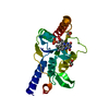
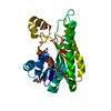
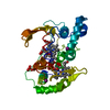
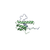
- PDB-3ebe: Crystal structure of xenopus laevis replication initiation factor... -
+ Open data

- Basic information
| Entry | Database: PDB / ID: 3ebe | ||||||
|---|---|---|---|---|---|---|---|
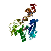
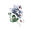
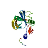
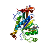
| Title | Crystal structure of xenopus laevis replication initiation factor MCM10 internal domain | ||||||
 Components Components | Protein MCM10 homolog | ||||||
 Keywords Keywords |  REPLICATION / OB-FOLD / ZINC FINGER / CCCH / DNA replication / Metal-binding / Nucleus / Zinc-finger REPLICATION / OB-FOLD / ZINC FINGER / CCCH / DNA replication / Metal-binding / Nucleus / Zinc-finger | ||||||
| Function / homology |  Function and homology information Function and homology informationDNA replication initiation / single-stranded DNA binding / double-stranded DNA binding / DNA damage response / metal ion binding / nucleusSimilarity search - Function | ||||||
| Biological species | Xenopus laevis (African clawed frog) | ||||||
| Method | X-RAY DIFFRACTION / SYNCHROTRON / MAD / Resolution: 2.3 Å | ||||||
 Authors Authors | Warren, E.M. / Eichman, B.F. | ||||||
 Citation Citation | Journal: Structure / Year: 2008 Title: Structural basis for DNA binding by replication initiator mcm10. Authors: Warren, E.M. / Vaithiyalingam, S. / Haworth, J. / Greer, B. / Bielinsky, A.K. / Chazin, W.J. / Eichman, B.F. | ||||||
| History |
|
- Structure visualization
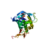
| Structure viewer | Molecule: MolmilJmol/JSmol |
|---|
- Downloads & links
-Download
| PDBx/mmCIF format | 3ebe.cif.gz | 117.6 KB | Display | PDBx/mmCIF format |
|---|---|---|---|---|
| PDB format | pdb3ebe.ent.gz | 91.9 KB | Display | PDB format |
| PDBx/mmJSON format | 3ebe.json.gz | Tree view | PDBx/mmJSON format | |
| Others |  Other downloads Other downloads |
-Validation report
| Arichive directory | https://data.pdbj.org/pub/pdb/validation_reports/eb/3ebeftp://data.pdbj.org/pub/pdb/validation_reports/eb/3ebe | HTTPS FTP |
|---|
-Related structure data
| Similar structure data |
|---|

-Links
 PDBj
PDBj
- Assembly
| Deposited unit | 
| ||||||||
|---|---|---|---|---|---|---|---|---|---|
| 1 | 
| ||||||||
| 2 | 
| ||||||||
| 3 | 
| ||||||||
| Unit cell |
|
-Components
| #1: Protein | Mass: 22699.984 Da / Num. of mol.: 3 / Fragment: UNP residues 230-427 Source method: isolated from a genetically manipulated source Source: (gene. exp.) Xenopus laevis (African clawed frog) / Gene: mcm10 / Production host:  Escherichia coli (E. coli) / Strain (production host): BL21(DE3) / References: UniProt: Q5EAW4 Escherichia coli (E. coli) / Strain (production host): BL21(DE3) / References: UniProt: Q5EAW4#2: Chemical |   Mass: 65.409 Da / Num. of mol.: 3 / Source method: obtained synthetically / Formula: Zn Mass: 65.409 Da / Num. of mol.: 3 / Source method: obtained synthetically / Formula: Zn#3: Water | ChemComp-HOH / | Water Mass: 18.015 Da / Num. of mol.: 149 / Source method: isolated from a natural source / Formula: H2O Mass: 18.015 Da / Num. of mol.: 149 / Source method: isolated from a natural source / Formula: H2O |
|---|
-Experimental details
-Experiment
| Experiment | Method: X-RAY DIFFRACTION / Number of used crystals: 2 |
|---|
- Sample preparation
| Crystal | Density Matthews: 2.55 Å3/Da / Density % sol: 51.84 % |
|---|---|
| Crystal grow | Temperature: 294 K / Method: vapor diffusion, hanging drop / pH: 8 Details: PEG 4000, TRIS, KSCN, pH 8.00, VAPOR DIFFUSION, HANGING DROP, temperature 294K |
-Data collection
| Diffraction |
| ||||||||||||||||||
|---|---|---|---|---|---|---|---|---|---|---|---|---|---|---|---|---|---|---|---|
| Diffraction source |
| ||||||||||||||||||
| Detector |
| ||||||||||||||||||
| Radiation |
| ||||||||||||||||||
| Radiation wavelength |
| ||||||||||||||||||
| Reflection | Resolution: 2.3→50 Å / Num. obs: 29404 / % possible obs: 99.6 % / Observed criterion σ(F): 0 / Observed criterion σ(I): 0 / Redundancy: 6.7 % / Biso Wilson estimate: 48.4 Å2 / Rsym value: 0.074 / Net I/σ(I): 27.7 | ||||||||||||||||||
| Reflection shell | Resolution: 2.3→2.38 Å / Redundancy: 3.9 % / Mean I/σ(I) obs: 2.3 / Num. unique all: 2882 / Rsym value: 0.425 / % possible all: 97 |

- Processing
| Software |
| ||||||||||||||||||||||||||||||||||||||||||||||||||||||||||||||||||||||||||||||||||||||||||||||||||||
|---|---|---|---|---|---|---|---|---|---|---|---|---|---|---|---|---|---|---|---|---|---|---|---|---|---|---|---|---|---|---|---|---|---|---|---|---|---|---|---|---|---|---|---|---|---|---|---|---|---|---|---|---|---|---|---|---|---|---|---|---|---|---|---|---|---|---|---|---|---|---|---|---|---|---|---|---|---|---|---|---|---|---|---|---|---|---|---|---|---|---|---|---|---|---|---|---|---|---|---|---|---|
| Refinement | Method to determine structure: MAD / Resolution: 2.3→44.4 Å / Cor.coef. Fo:Fc: 0.947 / Cor.coef. Fo:Fc free: 0.924 / SU B: 14.508 / SU ML: 0.178 / TLS residual ADP flag: LIKELY RESIDUAL / Cross valid method: THROUGHOUT / ESU R: 0.305 / ESU R Free: 0.229 / Stereochemistry target values: MAXIMUM LIKELIHOOD
| ||||||||||||||||||||||||||||||||||||||||||||||||||||||||||||||||||||||||||||||||||||||||||||||||||||
| Solvent computation | Ion probe radii: 0.8 Å / Shrinkage radii: 0.8 Å / VDW probe radii: 1.2 Å / Solvent model: BABINET MODEL WITH MASK | ||||||||||||||||||||||||||||||||||||||||||||||||||||||||||||||||||||||||||||||||||||||||||||||||||||
| Displacement parameters | Biso mean: 55.685 Å2
| ||||||||||||||||||||||||||||||||||||||||||||||||||||||||||||||||||||||||||||||||||||||||||||||||||||
| Refinement step | Cycle: LAST / Resolution: 2.3→44.4 Å
| ||||||||||||||||||||||||||||||||||||||||||||||||||||||||||||||||||||||||||||||||||||||||||||||||||||
| Refine LS restraints |
| ||||||||||||||||||||||||||||||||||||||||||||||||||||||||||||||||||||||||||||||||||||||||||||||||||||
| LS refinement shell | Resolution: 2.3→2.424 Å / Total num. of bins used: 10
| ||||||||||||||||||||||||||||||||||||||||||||||||||||||||||||||||||||||||||||||||||||||||||||||||||||
| Refinement TLS params. | Method: refined / Refine-ID: X-RAY DIFFRACTION
| ||||||||||||||||||||||||||||||||||||||||||||||||||||||||||||||||||||||||||||||||||||||||||||||||||||
| Refinement TLS group |
|
