 Movie
Movie Controller

[English] 日本語
 Yorodumi
- PDB-3dx3: Golgi alpha-Mannosidase II in complex with Mannostatin analog (1R... -
+ Open data

- Basic information
| Entry | Database: PDB / ID: 3dx3 | ||||||
|---|---|---|---|---|---|---|---|
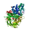
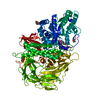
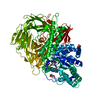
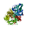
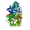
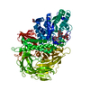
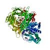
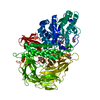
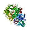
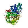
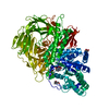
| Title | Golgi alpha-Mannosidase II in complex with Mannostatin analog (1R,2R,3S,4R,5R)-5-aminocyclopentane-1,2,3,4-tetraol | ||||||
 Components Components | Alpha-mannosidase 2 | ||||||
 Keywords Keywords | HYDROLASE / GH38 Glycosidase / Glycosidase / Golgi apparatus / Membrane / Metal-binding / Signal-anchor / Transmembrane | ||||||
| Function / homology |  Function and homology informationmannosyl-oligosaccharide 1,3-1,6-alpha-mannosidase / mannosyl-oligosaccharide 1,3-1,6-alpha-mannosidase activity / rhodopsin biosynthetic process / encapsulation of foreign target / Reactions specific to the complex N-glycan synthesis pathway / mannosidase activity / alpha-mannosidase activity / N-glycan processing / mannose metabolic process / Golgi stack ...mannosyl-oligosaccharide 1,3-1,6-alpha-mannosidase / mannosyl-oligosaccharide 1,3-1,6-alpha-mannosidase activity / rhodopsin biosynthetic process / encapsulation of foreign target / Reactions specific to the complex N-glycan synthesis pathway / mannosidase activity / alpha-mannosidase activity / N-glycan processing / mannose metabolic process / Golgi stack / protein deglycosylation / protein glycosylation / carbohydrate binding / Golgi membrane / endoplasmic reticulum / metal ion binding Function and homology informationmannosyl-oligosaccharide 1,3-1,6-alpha-mannosidase / mannosyl-oligosaccharide 1,3-1,6-alpha-mannosidase activity / rhodopsin biosynthetic process / encapsulation of foreign target / Reactions specific to the complex N-glycan synthesis pathway / mannosidase activity / alpha-mannosidase activity / N-glycan processing / mannose metabolic process / Golgi stack ...mannosyl-oligosaccharide 1,3-1,6-alpha-mannosidase / mannosyl-oligosaccharide 1,3-1,6-alpha-mannosidase activity / rhodopsin biosynthetic process / encapsulation of foreign target / Reactions specific to the complex N-glycan synthesis pathway / mannosidase activity / alpha-mannosidase activity / N-glycan processing / mannose metabolic process / Golgi stack / protein deglycosylation / protein glycosylation / carbohydrate binding / Golgi membrane / endoplasmic reticulum / metal ion bindingSimilarity search - Function | ||||||
| Biological species |  Drosophila melanogaster (fruit fly) Drosophila melanogaster (fruit fly) | ||||||
| Method | X-RAY DIFFRACTION / SYNCHROTRON / MOLECULAR REPLACEMENT / Resolution: 1.42 Å | ||||||
 Authors Authors | Kuntz, D.A. / Rose, D.R. | ||||||
 Citation Citation | Journal: Chembiochem / Year: 2009 Title: The molecular basis of inhibition of Golgi alpha-mannosidase II by mannostatin A. Authors: Kuntz, D.A. / Zhong, W. / Guo, J. / Rose, D.R. / Boons, G.J. | ||||||
| History |
|
- Structure visualization
| Structure viewer | Molecule: MolmilJmol/JSmol |
|---|
- Downloads & links
-Download
| PDBx/mmCIF format | 3dx3.cif.gz | 480 KB | Display | PDBx/mmCIF format |
|---|---|---|---|---|
| PDB format | pdb3dx3.ent.gz | 388 KB | Display | PDB format |
| PDBx/mmJSON format | 3dx3.json.gz | Tree view | PDBx/mmJSON format | |
| Others |  Other downloads Other downloads |
-Validation report
| Arichive directory | https://data.pdbj.org/pub/pdb/validation_reports/dx/3dx3ftp://data.pdbj.org/pub/pdb/validation_reports/dx/3dx3 | HTTPS FTP |
|---|
-Related structure data
| Related structure data |  3dx0C  3dx1C  3dx2C  3dx4C 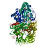 1htyS S: Starting model for refinement C: citing same article ( |
|---|---|
| Similar structure data |


-Links
 PDBj
PDBj


- Assembly
| Deposited unit | 
| ||||||||
|---|---|---|---|---|---|---|---|---|---|
| 1 |
| ||||||||
| Unit cell |
|
-Components
-Protein / Sugars , 2 types, 2 molecules A
| #1: Protein | / Alpha-mannosidase II / AMAN II / MAN II / Mannosyl-oligosaccharide 1 / 3-1 / 6-alpha-mannosidase / ...Alpha-mannosidase II / AMAN II / MAN II / Mannosyl-oligosaccharide 1 / 3-1 / 6-alpha-mannosidase / Golgi alpha-mannosidase II Mass: 119701.617 Da / Num. of mol.: 1 / Fragment: Catalytic domain; UNP residues 76-1108 Source method: isolated from a genetically manipulated source Source: (gene. exp.) Drosophila melanogaster (fruit fly) / Gene: alpha-Man-II, GmII, CG18802 / Plasmid: pMTBIP_NHIS / Production host: Drosophila melanogaster (fruit fly) / Strain (production host): S2 cellsReferences: UniProt: Q24451, mannosyl-oligosaccharide 1,3-1,6-alpha-mannosidase |
|---|---|
| #2: Sugar | ChemComp-NAG / N-Acetylglucosamine Type: D-saccharide, beta linking / Mass: 221.208 Da / Num. of mol.: 1 Type: D-saccharide, beta linking / Mass: 221.208 Da / Num. of mol.: 1Source method: isolated from a genetically manipulated source Formula: C8H15NO6 |
-Non-polymers , 5 types, 1403 molecules 


| #3: Chemical | ChemComp-ZN /  Mass: 65.409 Da / Num. of mol.: 1 / Source method: obtained synthetically / Formula: Zn Mass: 65.409 Da / Num. of mol.: 1 / Source method: obtained synthetically / Formula: Zn |
|---|---|
| #4: Chemical | ChemComp-PO4 / Phosphate Mass: 94.971 Da / Num. of mol.: 1 / Source method: obtained synthetically / Formula: PO4 Mass: 94.971 Da / Num. of mol.: 1 / Source method: obtained synthetically / Formula: PO4 |
| #5: Chemical | ChemComp-MRD / (2-Methyl-2,4-pentanediol Mass: 118.174 Da / Num. of mol.: 1 / Source method: obtained synthetically / Formula: C6H14O2 / Comment: precipitant*YM Mass: 118.174 Da / Num. of mol.: 1 / Source method: obtained synthetically / Formula: C6H14O2 / Comment: precipitant*YM |
| #6: Chemical | ChemComp-YTB / ( Mass: 149.145 Da / Num. of mol.: 1 / Source method: obtained synthetically / Formula: C5H11NO4 Mass: 149.145 Da / Num. of mol.: 1 / Source method: obtained synthetically / Formula: C5H11NO4 |
| #7: Water | ChemComp-HOH / WaterMass: 18.015 Da / Num. of mol.: 1399 / Source method: isolated from a natural source / Formula: H2O |
-Details
| Sequence details | E970K CONFLICT IN UNP ENTRY Q24451 |
|---|
-Experimental details
-Experiment
| Experiment | Method: X-RAY DIFFRACTION / Number of used crystals: 1 |
|---|
- Sample preparation
| Crystal | Density Matthews: 2.2 Å3/Da / Density % sol: 44.08 % |
|---|---|
| Crystal grow | Temperature: 298 K / Method: vapor diffusion, hanging drop / pH: 7 Details: PEG8000, Tris, 2.5% MPD, VAPOR DIFFUSION, HANGING DROP, temperature 298K, pH 7 |
-Data collection
| Diffraction | Mean temperature: 100 K |
|---|---|
| Diffraction source | Source: SYNCHROTRON / Site: CHESS  / Beamline: A1 / Wavelength: 0.977 Å / Beamline: A1 / Wavelength: 0.977 Å |
| Detector | Type: ADSC QUANTUM 4 / Detector: CCD / Date: Apr 20, 2007 |
| Radiation | Protocol: SINGLE WAVELENGTH / Monochromatic (M) / Laue (L): M / Scattering type: x-ray |
| Radiation wavelength | Wavelength: 0.977 Å / Relative weight: 1 |
| Reflection | Resolution: 1.42→40 Å / Num. obs: 193233 / % possible obs: 97.1 % / Observed criterion σ(F): 0 / Redundancy: 7.4 % / Rmerge(I) obs: 0.074 / Net I/σ(I): 18.2 |
| Reflection shell | Resolution: 1.42→1.46 Å / Redundancy: 5.1 % / Rmerge(I) obs: 0.26 / Mean I/σ(I) obs: 6.6 / Num. unique all: 11985 / % possible all: 78.8 |
- Processing
| Software |
| |||||||||||||||||||||||||||||||||||||||||||||||||||||||||||||||||||||||||||||||||||||||||||||||||||||||||
|---|---|---|---|---|---|---|---|---|---|---|---|---|---|---|---|---|---|---|---|---|---|---|---|---|---|---|---|---|---|---|---|---|---|---|---|---|---|---|---|---|---|---|---|---|---|---|---|---|---|---|---|---|---|---|---|---|---|---|---|---|---|---|---|---|---|---|---|---|---|---|---|---|---|---|---|---|---|---|---|---|---|---|---|---|---|---|---|---|---|---|---|---|---|---|---|---|---|---|---|---|---|---|---|---|---|---|
| Refinement | Method to determine structure: MOLECULAR REPLACEMENT Starting model: PDB entry 1hty Resolution: 1.42→19.52 Å / Cor.coef. Fo:Fc: 0.966 / Cor.coef. Fo:Fc free: 0.961 / Occupancy max: 1 / Occupancy min: 0.5 / FOM work R set: 0.906 / SU B: 1.733 / SU ML: 0.032 / Cross valid method: THROUGHOUT / σ(F): 0 / ESU R: 0.067 / ESU R Free: 0.057 / Stereochemistry target values: MAXIMUM LIKELIHOOD / Details: HYDROGENS HAVE BEEN ADDED IN THE RIDING POSITIONS
| |||||||||||||||||||||||||||||||||||||||||||||||||||||||||||||||||||||||||||||||||||||||||||||||||||||||||
| Solvent computation | Ion probe radii: 0.8 Å / Shrinkage radii: 0.8 Å / VDW probe radii: 1.2 Å / Solvent model: MASK | |||||||||||||||||||||||||||||||||||||||||||||||||||||||||||||||||||||||||||||||||||||||||||||||||||||||||
| Displacement parameters | Biso max: 59.24 Å2 / Biso mean: 14.421 Å2 / Biso min: 3.23 Å2
| |||||||||||||||||||||||||||||||||||||||||||||||||||||||||||||||||||||||||||||||||||||||||||||||||||||||||
| Refinement step | Cycle: LAST / Resolution: 1.42→19.52 Å
| |||||||||||||||||||||||||||||||||||||||||||||||||||||||||||||||||||||||||||||||||||||||||||||||||||||||||
| Refine LS restraints |
| |||||||||||||||||||||||||||||||||||||||||||||||||||||||||||||||||||||||||||||||||||||||||||||||||||||||||
| LS refinement shell | Resolution: 1.42→1.457 Å / Total num. of bins used: 20
|
