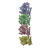
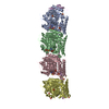
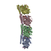
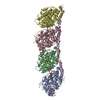
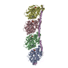
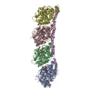
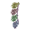
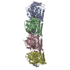
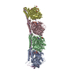
THERE IS ONE COMPLEX IN THE ASYMMETRIC UNIT, WHICH CONSISTS OF TWO ALPHA-BETA TUBULIN HETERODIMERS ...THERE IS ONE COMPLEX IN THE ASYMMETRIC UNIT, WHICH CONSISTS OF TWO ALPHA-BETA TUBULIN HETERODIMERS (CHAINS A-B AND C-D), AND ONE STATHMIN-LIKE DOMAIN OF RB3 (RB3-SLD) WHICH CORRESPONDS TO STAHMIN RESIDUES 5 TO 145 WITH THE ADDITION OF ONE ACETYLATED ALANINE AT THE N-TERMINUS. THE NUMBERING OF RB3-SLD IS ACCORDING TO THE STATHMIN SEQUENCE. ALPHA- TUBULIN AND BETA-TUBULIN HAVE BEEN ALIGNED AS IN NOGALES ET AL., NATURE VOL 391,199-203. IN THIS ALIGNMENT, RESIDUES 45-46 AND 361-368 OF ALPHA-TUBULIN ARE MISSING IN BETA- TUBULIN. THERE ARE SEVERAL EXPRESSED TUBULIN ISOTYPES IN MAMMALIAN BRAIN. WE USED THE BOS TAURUS TUBULIN SEQUENCE OF THE MOST ABUNDANT ISOTYPES (ALPHA: ISOTYPE 1, GI:73586894, BETA: ISOTYPE 2, GI:51491829) BUT WITH THE ILE TO VAL SUBSTITUTION AT POSITION 318 ON BETA TUBULIN.
-
Experimental details
-
Experiment
Experiment
Method: X-RAY DIFFRACTION / Number of used crystals: 1
-
Sample preparation
Crystal
Density Matthews: 3.82 Å3/Da / Density % sol: 68 %
Crystal grow
Temperature: 291 K / Method: vapor diffusion, hanging drop / pH: 7 Details: PEG, PIPES BUFFER. The crystal of tubulin-colchicine:RB3-SLD complex was soaked with a 0.7mM Phomopsin A solution for 24 hours., pH 7.00, VAPOR DIFFUSION, HANGING DROP, temperature 291K
Resolution: 4.1→20 Å / Cor.coef. Fo:Fc: 0.955 / Cor.coef. Fo:Fc free: 0.938 / SU B: 65.918 / SU ML: 0.784 / TLS residual ADP flag: LIKELY RESIDUAL / Cross valid method: THROUGHOUT / σ(F): 0 / ESU R Free: 0.86 / Stereochemistry target values: MAXIMUM LIKELIHOOD Details: Care should be exercised in interpreting the current model due to the limited (4.1 angstroms) resolution.
Rfactor
Num. reflection
% reflection
Selection details
Rfree
0.26471
1277
5 %
RANDOM
Rwork
0.21521
-
-
-
all
0.21781
26330
-
-
obs
0.21781
25389
96.43 %
-
Solvent computation
Ion probe radii: 0.8 Å / Shrinkage radii: 0.8 Å / VDW probe radii: 1.4 Å / Solvent model: BABINET MODEL WITH MASK
Displacement parameters
Biso mean: 95.778 Å2
Baniso -1
Baniso -2
Baniso -3
1-
3.55 Å2
1.77 Å2
0 Å2
2-
-
3.55 Å2
0 Å2
3-
-
-
-5.32 Å2
Refinement step
Cycle: LAST / Resolution: 4.1→20 Å
Protein
Nucleic acid
Ligand
Solvent
Total
Num. atoms
13966
0
292
0
14258
Refine LS restraints
Refine-ID
Type
Dev ideal
Dev ideal target
Number
X-RAY DIFFRACTION
r_bond_refined_d
0.017
0.021
14583
X-RAY DIFFRACTION
r_angle_refined_deg
2.041
1.96
19873
X-RAY DIFFRACTION
r_dihedral_angle_1_deg
9.774
5
1833
X-RAY DIFFRACTION
r_chiral_restr
0.131
0.2
2185
X-RAY DIFFRACTION
r_gen_planes_refined
0.005
0.02
11309
X-RAY DIFFRACTION
r_nbd_refined
0.305
0.2
8101
X-RAY DIFFRACTION
r_xyhbond_nbd_refined
0.241
0.2
648
X-RAY DIFFRACTION
r_metal_ion_refined
0.574
0.2
4
X-RAY DIFFRACTION
r_symmetry_vdw_refined
0.385
0.2
47
X-RAY DIFFRACTION
r_symmetry_hbond_refined
0.449
0.2
2
X-RAY DIFFRACTION
r_mcbond_it
0
1.5
9145
X-RAY DIFFRACTION
r_mcangle_it
0
2
14597
X-RAY DIFFRACTION
r_scbond_it
0
3
5438
X-RAY DIFFRACTION
r_scangle_it
0
4.5
5275
Refine LS restraints NCS
Dom-ID: 1 / Refine-ID: X-RAY DIFFRACTION
Ens-ID
Auth asym-ID
Number
Type
Rms dev position (Å)
Weight position
1
A
3114
tightpositional
0.07
0.05
2
B
3170
tightpositional
0.06
0.05
1
A
3114
tightthermal
0
0.5
2
B
3170
tightthermal
0
0.5
LS refinement shell
Resolution: 4.1→4.202 Å / Total num. of bins used: 20 /
Rfactor
Num. reflection
Rfree
0.457
85
Rwork
0.415
1579
Refinement TLS params.
Method: refined / Refine-ID: X-RAY DIFFRACTION
ID
L11 (°2)
L12 (°2)
L13 (°2)
L22 (°2)
L23 (°2)
L33 (°2)
S11 (Å °)
S12 (Å °)
S13 (Å °)
S21 (Å °)
S22 (Å °)
S23 (Å °)
S31 (Å °)
S32 (Å °)
S33 (Å °)
T11 (Å2)
T12 (Å2)
T13 (Å2)
T22 (Å2)
T23 (Å2)
T33 (Å2)
Origin x (Å)
Origin y (Å)
Origin z (Å)
1
9.31
0.7414
0.4089
3.7397
-0.2398
2.7307
0.3313
-0.3905
0.8792
0.1902
-0.1885
0.0397
-0.4371
0.0717
-0.1428
0.7402
-0.2375
0.1502
0.8799
0.1293
1.3701
134.965
105.038
16.588
2
8.1176
1.8013
0.3755
4.9741
-0.2661
4.6014
0.3047
-0.1748
-0.518
-0.1036
-0.2085
-0.2275
0.2506
0.3135
-0.0963
0.6614
-0.2581
0.0079
1.2136
0.2259
1.0843
102.16
80.932
4.468
3
10.5655
2.9539
-1.044
4.2999
0.2576
3.4598
0.2206
0.256
-0.6378
-0.3317
0.1518
-0.137
0.0307
0.2211
-0.3724
0.8047
-0.2032
-0.4189
1.4182
0.416
1.5655
63.859
60.791
-2.668
4
8.792
2.9771
-0.0922
5.5153
0.6811
8.6008
0.317
0.5166
0.259
0.398
-0.8714
0.7603
0.575
-0.6993
0.5544
0.3653
-0.0833
-0.6826
1.8522
0.1543
1.2757
24.485
48.231
-5.809
Refinement TLS group
ID
Refine-ID
Refine TLS-ID
Auth asym-ID
Label asym-ID
Auth seq-ID
Label seq-ID
1
X-RAY DIFFRACTION
1
A
A
2 - 439
2 - 439
2
X-RAY DIFFRACTION
1
E
E
4 - 64
1 - 61
3
X-RAY DIFFRACTION
1
A
F
500
1
4
X-RAY DIFFRACTION
1
D
H
600
1
5
X-RAY DIFFRACTION
2
B
B
2 - 437
2 - 427
6
X-RAY DIFFRACTION
2
E
E
65 - 89
62 - 86
7
X-RAY DIFFRACTION
2
D
H
602
3
8
X-RAY DIFFRACTION
2
B
I
700
1
9
X-RAY DIFFRACTION
2
B
K
800
1
10
X-RAY DIFFRACTION
3
C
C
2 - 439
2 - 439
11
X-RAY DIFFRACTION
3
E
E
90 - 115
87 - 112
12
X-RAY DIFFRACTION
3
C
G
501
1
13
X-RAY DIFFRACTION
3
D
H
601
2
14
X-RAY DIFFRACTION
4
D
D
2 - 437
2 - 427
15
X-RAY DIFFRACTION
4
E
E
116 - 141
113 - 138
16
X-RAY DIFFRACTION
4
D
H
603
4
17
X-RAY DIFFRACTION
4
D
J
701
1
18
X-RAY DIFFRACTION
4
D
L
801
1
+
About Yorodumi
-
News
-
Feb 9, 2022. New format data for meta-information of EMDB entries
New format data for meta-information of EMDB entries
Version 3 of the EMDB header file is now the official format.
The previous official version 1.9 will be removed from the archive.
In the structure databanks used in Yorodumi, some data are registered as the other names, "COVID-19 virus" and "2019-nCoV". Here are the details of the virus and the list of structure data.
Jan 31, 2019. EMDB accession codes are about to change! (news from PDBe EMDB page)
EMDB accession codes are about to change! (news from PDBe EMDB page)
The allocation of 4 digits for EMDB accession codes will soon come to an end. Whilst these codes will remain in use, new EMDB accession codes will include an additional digit and will expand incrementally as the available range of codes is exhausted. The current 4-digit format prefixed with “EMD-” (i.e. EMD-XXXX) will advance to a 5-digit format (i.e. EMD-XXXXX), and so on. It is currently estimated that the 4-digit codes will be depleted around Spring 2019, at which point the 5-digit format will come into force.
The EM Navigator/Yorodumi systems omit the EMD- prefix.
Related info.:Q: What is EMD? / ID/Accession-code notation in Yorodumi/EM Navigator
Yorodumi is a browser for structure data from EMDB, PDB, SASBDB, etc.
This page is also the successor to EM Navigator detail page, and also detail information page/front-end page for Omokage search.
The word "yorodu" (or yorozu) is an old Japanese word meaning "ten thousand". "mi" (miru) is to see.
Related info.:EMDB / PDB / SASBDB / Comparison of 3 databanks / Yorodumi Search / Aug 31, 2016. New EM Navigator & Yorodumi / Yorodumi Papers / Jmol/JSmol / Function and homology information / Changes in new EM Navigator and Yorodumi
 Movie
Movie Controller


 Open data
 Basic information
 Components
 Keywords
 CELL CYCLE / alpha-tubulin /
  Function and homology information
 Authors
 Citation
 Structure visualization
 Downloads & links
 Other downloads


 PDBj
PDBj


 Assembly
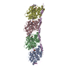


 Mass: 24.305 Da / Num. of mol.: 2 / Source method: obtained synthetically / Formula: Mg
 Mass: 431.502 Da / Num. of mol.: 2 / Source method: obtained synthetically / Formula: C22H25NO6S
 Mass: 789.228 Da / Num. of mol.: 2 / Source method: obtained synthetically / Formula: C36H45ClN6O12
 Mass: 523.180 Da / Num. of mol.: 2 / Source method: obtained synthetically / Formula: C10H16N5O14P3 / Comment: GTP, energy-carrying molecule*YM
 Type: RNA linking / Mass: 443.201 Da / Num. of mol.: 2 / Source method: obtained synthetically / Formula: C10H15N5O11P2 / Comment: GDP, energy-carrying molecule*YM
 Sample preparation
 / Beamline: ID14-4 / Wavelength: 0.976 / Wavelength: 0.976 Å
 Processing