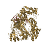
 Movie
Movie Controller

[English] 日本語
 Yorodumi
- PDB-3dky: Crystal Structure of the replication initiator protein encoded on... -
+ Open data

- Basic information
| Entry | Database: PDB / ID: 3dky | ||||||
|---|---|---|---|---|---|---|---|
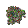
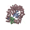
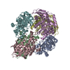
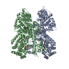
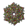
| Title | Crystal Structure of the replication initiator protein encoded on plasmid pMV158 (RepB), tetragonal form, to 3.6 Ang resolution | ||||||
 Components Components | Replication protein repB DNA replication DNA replication | ||||||
 Keywords Keywords | REPLICATION / Replication initiation / Plasmid replication / Nuclease / Hexamer / Flexible nuclease domains / DNA replication / Plasmid / REPLICATION INITIATOR | ||||||
| Function / homology |  Function and homology informationDNA topoisomerase activity / extrachromosomal circular DNA / DNA replication / DNA binding / identical protein binding Function and homology informationDNA topoisomerase activity / extrachromosomal circular DNA / DNA replication / DNA binding / identical protein bindingSimilarity search - Function | ||||||
| Biological species |  Streptococcus agalactiae (bacteria) Streptococcus agalactiae (bacteria) | ||||||
| Method | X-RAY DIFFRACTION / SYNCHROTRON / MOLECULAR REPLACEMENT / Resolution: 3.6 Å | ||||||
 Authors Authors | Boer, D.R. / Ruiz-Maso, J.A. / Blanco, A.G. / Vives-Llacer, M. / Uson, I. / Gomis-Ruth, F.X. / Espinosa, M. / Del Solar, G. / Coll, M. | ||||||
 Citation Citation | Journal: Embo J. / Year: 2009 Title: Plasmid replication initiator RepB forms a hexamer reminiscent of ring helicases and has mobile nuclease domains Authors: Boer, D.R. / Ruiz-Maso, J.A. / Lopez-Blanco, J.R. / Blanco, A.G. / Vives-Llacer, M. / Chacon, P. / Uson, I. / Gomis-Ruth, F.X. / Espinosa, M. / Llorca, O. / del Solar, G. / Coll, M. | ||||||
| History |
|
- Structure visualization
| Structure viewer | Molecule: MolmilJmol/JSmol |
|---|
- Downloads & links
-Download
| PDBx/mmCIF format | 3dky.cif.gz | 487.8 KB | Display | PDBx/mmCIF format |
|---|---|---|---|---|
| PDB format | pdb3dky.ent.gz | 411.7 KB | Display | PDB format |
| PDBx/mmJSON format | 3dky.json.gz | Tree view | PDBx/mmJSON format | |
| Others |  Other downloads Other downloads |
-Validation report
| Arichive directory | https://data.pdbj.org/pub/pdb/validation_reports/dk/3dkyftp://data.pdbj.org/pub/pdb/validation_reports/dk/3dky | HTTPS FTP |
|---|
-Related structure data
| Related structure data |  3dkxSC S: Starting model for refinement C: citing same article ( |
|---|---|
| Similar structure data |


-Links
 PDBj
PDBj- Assembly
| Deposited unit | 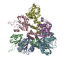
| |||||||||||||||||||||||||||||||||||||||||||||||||||||||||||||||||||||||||||||||||||||||||||||||||||||||||||||||||||||||||||||||||||||||||||
|---|---|---|---|---|---|---|---|---|---|---|---|---|---|---|---|---|---|---|---|---|---|---|---|---|---|---|---|---|---|---|---|---|---|---|---|---|---|---|---|---|---|---|---|---|---|---|---|---|---|---|---|---|---|---|---|---|---|---|---|---|---|---|---|---|---|---|---|---|---|---|---|---|---|---|---|---|---|---|---|---|---|---|---|---|---|---|---|---|---|---|---|---|---|---|---|---|---|---|---|---|---|---|---|---|---|---|---|---|---|---|---|---|---|---|---|---|---|---|---|---|---|---|---|---|---|---|---|---|---|---|---|---|---|---|---|---|---|---|---|---|
| 1 |
| |||||||||||||||||||||||||||||||||||||||||||||||||||||||||||||||||||||||||||||||||||||||||||||||||||||||||||||||||||||||||||||||||||||||||||
| Unit cell |
| |||||||||||||||||||||||||||||||||||||||||||||||||||||||||||||||||||||||||||||||||||||||||||||||||||||||||||||||||||||||||||||||||||||||||||
| Noncrystallographic symmetry (NCS) | NCS domain:
NCS domain segments:
|