 Movie
Movie Controller


+ Open data

- Basic information
| Entry | Database: PDB / ID: 3djt | ||||||
|---|---|---|---|---|---|---|---|
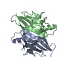
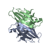
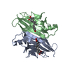
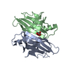
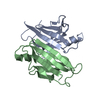
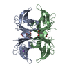
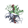
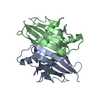
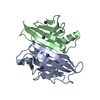
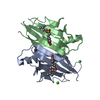
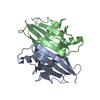
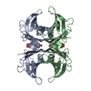
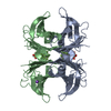
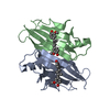
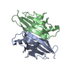
| Title | Crystal structure of transthyretin variant V30M at acidic pH | ||||||
 Components Components | Transthyretin | ||||||
 Keywords Keywords | TRANSPORT PROTEIN / TTR / AMYLOID FIBRILS / POINT MUTATION / Amyloid / Disease mutation / Gamma-carboxyglutamic acid / Glycoprotein / Hormone / Polyneuropathy / Retinol-binding / Secreted / Thyroid hormone / Transport / Vitamin A | ||||||
| Function / homology |  Function and homology information Function and homology informationRetinoid cycle disease events / thyroid hormone binding / The canonical retinoid cycle in rods (twilight vision) / Non-integrin membrane-ECM interactions / purine nucleobase metabolic process / Retinoid metabolism and transport / hormone activity / azurophil granule lumen / Amyloid fiber formation / Neutrophil degranulation ...Retinoid cycle disease events / thyroid hormone binding / The canonical retinoid cycle in rods (twilight vision) / Non-integrin membrane-ECM interactions / purine nucleobase metabolic process / Retinoid metabolism and transport / hormone activity / azurophil granule lumen / Amyloid fiber formation / Neutrophil degranulation / extracellular space / extracellular exosome / extracellular region / identical protein bindingSimilarity search - Function | ||||||
| Biological species |  HOMO SAPIENS (human) HOMO SAPIENS (human) | ||||||
| Method | X-RAY DIFFRACTION / SYNCHROTRON / MOLECULAR REPLACEMENT / Resolution: 2.3 Å | ||||||
 Authors Authors | Cendron, L. / Zanotti, G. / Folli, C. / Berni, R. | ||||||
 Citation Citation | Journal: J.Biol.Chem. / Year: 2009 Title: Amyloidogenic potential of transthyretin variants: insights from structural and computational analyses. Authors: Cendron, L. / Trovato, A. / Seno, F. / Folli, C. / Alfieri, B. / Zanotti, G. / Berni, R. #1: Journal: J.Mol.Biol. / Year: 2007Title: Acidic Ph-Induced Conformational Changes in Amyloidogenic Mutant Transthyretin. Authors: Pasquato, N. / Berni, R. / Folli, C. / Alfieri, B. / Cendron, L. / Zanotti, G. #2: Journal: J.Mol.Biol. / Year: 2000Title: A Comparative Analysis of 23 Structures of the Amyloidogenic Protein Transthyretin. Authors: Eneqvist, T. / Olofsson, A. / Lundgren, E. / Sauer-Eriksson, A.E. | ||||||
| History |
|
- Structure visualization
| Structure viewer | Molecule: MolmilJmol/JSmol |
|---|
- Downloads & links
-Download
| PDBx/mmCIF format | 3djt.cif.gz | 60.5 KB | Display | PDBx/mmCIF format |
|---|---|---|---|---|
| PDB format | pdb3djt.ent.gz | 45.2 KB | Display | PDB format |
| PDBx/mmJSON format | 3djt.json.gz | Tree view | PDBx/mmJSON format | |
| Others |  Other downloads Other downloads |
-Validation report
| Arichive directory | https://data.pdbj.org/pub/pdb/validation_reports/dj/3djtftp://data.pdbj.org/pub/pdb/validation_reports/dj/3djt | HTTPS FTP |
|---|
-Related structure data
| Related structure data |  3djrC  3djsC  3djzC  3dk0C  3dk2C  1f41S S: Starting model for refinement C: citing same article ( |
|---|---|
| Similar structure data |


-Links
 PDBj
PDBj


- Assembly
| Deposited unit | 
| ||||||||||||
|---|---|---|---|---|---|---|---|---|---|---|---|---|---|
| 1 | 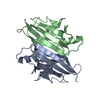
| ||||||||||||
| Unit cell |
| ||||||||||||
| Components on special symmetry positions |
|
-Components
| #1: Protein | / Prealbumin / TBPA / TTR / ATTR Mass: 13809.426 Da / Num. of mol.: 2 / Mutation: V30M Source method: isolated from a genetically manipulated source Source: (gene. exp.) HOMO SAPIENS (human) / Production host:  ESCHERICHIA COLI (E. coli) / Strain (production host): BL21 (DE3) / References: UniProt: P02766 ESCHERICHIA COLI (E. coli) / Strain (production host): BL21 (DE3) / References: UniProt: P02766#2: Water | ChemComp-HOH / | Water Mass: 18.015 Da / Num. of mol.: 136 / Source method: isolated from a natural source / Formula: H2O Mass: 18.015 Da / Num. of mol.: 136 / Source method: isolated from a natural source / Formula: H2O |
|---|
-Experimental details
-Experiment
| Experiment | Method: X-RAY DIFFRACTION / Number of used crystals: 1 |
|---|
- Sample preparation
| Crystal | Density Matthews: 2.19 Å3/Da / Density % sol: 43.85 % |
|---|---|
| Crystal grow | Temperature: 295 K / Method: vapor diffusion / pH: 4.6 Details: 32% PEG 2000, 0.1M NaAc, 0.2M Am2SO4 , pH 4.6, VAPOR DIFFUSION, temperature 295K |
-Data collection
| Diffraction | Mean temperature: 100 K |
|---|---|
| Diffraction source | Source: SYNCHROTRON / Site: ESRF  / Beamline: ID14-4 / Wavelength: 0.9859 Å / Beamline: ID14-4 / Wavelength: 0.9859 Å |
| Detector | Type: ADSC QUANTUM 315 / Detector: CCD / Date: Dec 7, 2006 |
| Radiation | Protocol: SINGLE WAVELENGTH / Monochromatic (M) / Laue (L): M / Scattering type: x-ray |
| Radiation wavelength | Wavelength: 0.9859 Å / Relative weight: 1 |
| Reflection | Resolution: 2.2→86.07 Å / Num. all: 12491 / Num. obs: 12491 / % possible obs: 97.6 % / Observed criterion σ(F): 0 / Observed criterion σ(I): 0 / Redundancy: 3.3 % / Rmerge(I) obs: 0.094 / Net I/σ(I): 12 |
| Reflection shell | Resolution: 2.2→2.32 Å / Redundancy: 3 % / Rmerge(I) obs: 0.326 / Mean I/σ(I) obs: 5.5 / Num. unique all: 1716 / % possible all: 94.9 |
- Processing
| Software |
| ||||||||||||||||||||||||||||||||||||||||||||||||||||||||||||||||||||||||||||||||||||||||||||||||||||||||||||||||||||||||||||||||||||||||||||||||||||||||||||||||||||||||||
|---|---|---|---|---|---|---|---|---|---|---|---|---|---|---|---|---|---|---|---|---|---|---|---|---|---|---|---|---|---|---|---|---|---|---|---|---|---|---|---|---|---|---|---|---|---|---|---|---|---|---|---|---|---|---|---|---|---|---|---|---|---|---|---|---|---|---|---|---|---|---|---|---|---|---|---|---|---|---|---|---|---|---|---|---|---|---|---|---|---|---|---|---|---|---|---|---|---|---|---|---|---|---|---|---|---|---|---|---|---|---|---|---|---|---|---|---|---|---|---|---|---|---|---|---|---|---|---|---|---|---|---|---|---|---|---|---|---|---|---|---|---|---|---|---|---|---|---|---|---|---|---|---|---|---|---|---|---|---|---|---|---|---|---|---|---|---|---|---|---|---|---|
| Refinement | Method to determine structure: MOLECULAR REPLACEMENT Starting model: 1F41 Resolution: 2.3→51.5 Å / Cor.coef. Fo:Fc: 0.934 / Cor.coef. Fo:Fc free: 0.908 / SU B: 6.506 / SU ML: 0.162 / Cross valid method: THROUGHOUT / σ(F): 0 / σ(I): 1 / ESU R: 0.449 / ESU R Free: 0.26 / Stereochemistry target values: MAXIMUM LIKELIHOOD
| ||||||||||||||||||||||||||||||||||||||||||||||||||||||||||||||||||||||||||||||||||||||||||||||||||||||||||||||||||||||||||||||||||||||||||||||||||||||||||||||||||||||||||
| Solvent computation | Ion probe radii: 0.8 Å / Shrinkage radii: 0.8 Å / VDW probe radii: 1.2 Å / Solvent model: MASK | ||||||||||||||||||||||||||||||||||||||||||||||||||||||||||||||||||||||||||||||||||||||||||||||||||||||||||||||||||||||||||||||||||||||||||||||||||||||||||||||||||||||||||
| Displacement parameters | Biso mean: 31.92 Å2
| ||||||||||||||||||||||||||||||||||||||||||||||||||||||||||||||||||||||||||||||||||||||||||||||||||||||||||||||||||||||||||||||||||||||||||||||||||||||||||||||||||||||||||
| Refinement step | Cycle: LAST / Resolution: 2.3→51.5 Å
| ||||||||||||||||||||||||||||||||||||||||||||||||||||||||||||||||||||||||||||||||||||||||||||||||||||||||||||||||||||||||||||||||||||||||||||||||||||||||||||||||||||||||||
| Refine LS restraints |
| ||||||||||||||||||||||||||||||||||||||||||||||||||||||||||||||||||||||||||||||||||||||||||||||||||||||||||||||||||||||||||||||||||||||||||||||||||||||||||||||||||||||||||
| LS refinement shell | Resolution: 2.3→2.357 Å / Total num. of bins used: 20
|
