 Movie
Movie Controller


+ Open data

- Basic information
| Entry | Database: PDB / ID: 3d45 | ||||||
|---|---|---|---|---|---|---|---|
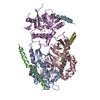
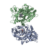
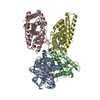
| Title | Crystal structure of mouse PARN in complex with m7GpppG | ||||||
 Components Components | Poly(A)-specific ribonuclease PARN | ||||||
 Keywords Keywords |  HYDROLASE / PARN / cap analogue / Exonuclease / Magnesium / Metal-binding / Nonsense-mediated mRNA decay / Nuclease / Nucleus / Phosphoprotein / RNA-binding HYDROLASE / PARN / cap analogue / Exonuclease / Magnesium / Metal-binding / Nonsense-mediated mRNA decay / Nuclease / Nucleus / Phosphoprotein / RNA-binding | ||||||
| Function / homology |  Function and homology information Function and homology informationKSRP (KHSRP) binds and destabilizes mRNA / : / box H/ACA sno(s)RNA 3'-end processing / Deadenylation of mRNA / poly(A)-specific ribonuclease / telomerase RNA stabilization / poly(A)-specific ribonuclease activity / cation binding / miRNA catabolic process / regulation of telomerase RNA localization to Cajal body ...KSRP (KHSRP) binds and destabilizes mRNA / : / box H/ACA sno(s)RNA 3'-end processing / Deadenylation of mRNA / poly(A)-specific ribonuclease / telomerase RNA stabilization / poly(A)-specific ribonuclease activity / cation binding / miRNA catabolic process / regulation of telomerase RNA localization to Cajal body / poly(A)-dependent snoRNA 3'-end processing / nuclear-transcribed mRNA poly(A) tail shortening / nuclear-transcribed mRNA catabolic process, nonsense-mediated decay / positive regulation of telomere maintenance via telomerase / postsynapse / nuclear speck / glutamatergic synapse / nucleolus / protein kinase binding / RNA binding / metal ion binding / cytoplasmSimilarity search - Function | ||||||
| Biological species |  Mus musculus (house mouse) Mus musculus (house mouse) | ||||||
| Method | X-RAY DIFFRACTION / SYNCHROTRON / MOLECULAR REPLACEMENT / Resolution: 3 Å | ||||||
 Authors Authors | Wu, M. / Song, H. | ||||||
 Citation Citation | Journal: Structure / Year: 2009 Title: Structural basis of m(7)GpppG binding to poly(A)-specific ribonuclease. Authors: Wu, M. / Nilsson, P. / Henriksson, N. / Niedzwiecka, A. / Lim, M.K. / Cheng, Z. / Kokkoris, K. / Virtanen, A. / Song, H. | ||||||
| History |
|
- Structure visualization
| Structure viewer | Molecule: MolmilJmol/JSmol |
|---|
- Downloads & links
-Download
| PDBx/mmCIF format | 3d45.cif.gz | 171.3 KB | Display | PDBx/mmCIF format |
|---|---|---|---|---|
| PDB format | pdb3d45.ent.gz | 132.3 KB | Display | PDB format |
| PDBx/mmJSON format | 3d45.json.gz | Tree view | PDBx/mmJSON format | |
| Others |  Other downloads Other downloads |
-Validation report
| Arichive directory | https://data.pdbj.org/pub/pdb/validation_reports/d4/3d45ftp://data.pdbj.org/pub/pdb/validation_reports/d4/3d45 | HTTPS FTP |
|---|
-Related structure data


-Links
 PDBj
PDBj

- Assembly
| Deposited unit | 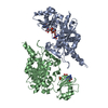
| |||||||||||||||||||||||||||||||||||||||||||||||||||||||||||||||||||||||||||||||||||||||||||||||||||||||||||||||||||||
|---|---|---|---|---|---|---|---|---|---|---|---|---|---|---|---|---|---|---|---|---|---|---|---|---|---|---|---|---|---|---|---|---|---|---|---|---|---|---|---|---|---|---|---|---|---|---|---|---|---|---|---|---|---|---|---|---|---|---|---|---|---|---|---|---|---|---|---|---|---|---|---|---|---|---|---|---|---|---|---|---|---|---|---|---|---|---|---|---|---|---|---|---|---|---|---|---|---|---|---|---|---|---|---|---|---|---|---|---|---|---|---|---|---|---|---|---|---|---|
| 1 |
| |||||||||||||||||||||||||||||||||||||||||||||||||||||||||||||||||||||||||||||||||||||||||||||||||||||||||||||||||||||
| Unit cell |
| |||||||||||||||||||||||||||||||||||||||||||||||||||||||||||||||||||||||||||||||||||||||||||||||||||||||||||||||||||||
| Noncrystallographic symmetry (NCS) | NCS domain:
NCS domain segments: Ens-ID: 1 / Refine code: 2
|
-Components
| #1: Protein | Mass: 58360.035 Da / Num. of mol.: 2 / Fragment: mPARN Source method: isolated from a genetically manipulated source Source: (gene. exp.) Mus musculus (house mouse) / Gene: Parn / Plasmid: pET28a / Production host:  Escherichia coli (E. coli) / Strain (production host): Bl-21 / References: UniProt: Q8VDG3, poly(A)-specific ribonuclease Escherichia coli (E. coli) / Strain (production host): Bl-21 / References: UniProt: Q8VDG3, poly(A)-specific ribonuclease#2: Chemical |   Type: RNA linking / Mass: 379.263 Da / Num. of mol.: 2 / Source method: obtained synthetically / Formula: C11H18N5O8P Type: RNA linking / Mass: 379.263 Da / Num. of mol.: 2 / Source method: obtained synthetically / Formula: C11H18N5O8P#3: Chemical | Guanosine diphosphate  Type: RNA linking / Mass: 443.201 Da / Num. of mol.: 2 / Source method: obtained synthetically / Formula: C10H15N5O11P2 / Comment: GDP, energy-carrying molecule*YM Type: RNA linking / Mass: 443.201 Da / Num. of mol.: 2 / Source method: obtained synthetically / Formula: C10H15N5O11P2 / Comment: GDP, energy-carrying molecule*YM#4: Water | ChemComp-HOH / | Water Mass: 18.015 Da / Num. of mol.: 78 / Source method: isolated from a natural source / Formula: H2O Mass: 18.015 Da / Num. of mol.: 78 / Source method: isolated from a natural source / Formula: H2O |
|---|
-Experimental details
-Experiment
| Experiment | Method: X-RAY DIFFRACTION / Number of used crystals: 1 |
|---|
- Sample preparation
| Crystal | Density Matthews: 2.82 Å3/Da / Density % sol: 56.39 % |
|---|---|
| Crystal grow | Temperature: 293 K / Method: vapor diffusion, hanging drop / pH: 6 Details: 6~10% PEG6000, 100mM MES, pH6.0, 10mM betaine , VAPOR DIFFUSION, HANGING DROP, temperature 293K |
-Data collection
| Diffraction | Mean temperature: 100 K |
|---|---|
| Diffraction source | Source: SYNCHROTRON / Site: ESRF  / Beamline: BM14 / Wavelength: 0.9793 Å / Beamline: BM14 / Wavelength: 0.9793 Å |
| Detector | Type: ADSC QUANTUM 210 / Detector: CCD |
| Radiation | Protocol: SINGLE WAVELENGTH / Monochromatic (M) / Laue (L): M / Scattering type: x-ray |
| Radiation wavelength | Wavelength: 0.9793 Å / Relative weight: 1 |
| Reflection | Resolution: 3→103.69 Å / Num. all: 29592 / Num. obs: 29592 / % possible obs: 96 % / Redundancy: 6.9 % / Rmerge(I) obs: 0.101 |
| Reflection shell | Resolution: 3→3.1 Å / Redundancy: 5.5 % / Rmerge(I) obs: 0.327 / Mean I/σ(I) obs: 2.3 / % possible all: 96 |
- Processing
| Software |
| ||||||||||||||||||||||||||||||||||||||||||||||||||||||||||||||||||||||||||||||||||||||||||
|---|---|---|---|---|---|---|---|---|---|---|---|---|---|---|---|---|---|---|---|---|---|---|---|---|---|---|---|---|---|---|---|---|---|---|---|---|---|---|---|---|---|---|---|---|---|---|---|---|---|---|---|---|---|---|---|---|---|---|---|---|---|---|---|---|---|---|---|---|---|---|---|---|---|---|---|---|---|---|---|---|---|---|---|---|---|---|---|---|---|---|---|
| Refinement | Method to determine structure: MOLECULAR REPLACEMENT Starting model: 2A1R 1WHV Resolution: 3→103.69 Å / Cor.coef. Fo:Fc: 0.835 / Cor.coef. Fo:Fc free: 0.838 / SU B: 40.73 / SU ML: 0.438 / TLS residual ADP flag: LIKELY RESIDUAL / Cross valid method: THROUGHOUT / ESU R Free: 0.526 / Stereochemistry target values: MAXIMUM LIKELIHOOD / Details: HYDROGENS HAVE BEEN ADDED IN THE RIDING POSITIONS
| ||||||||||||||||||||||||||||||||||||||||||||||||||||||||||||||||||||||||||||||||||||||||||
| Solvent computation | Ion probe radii: 0.8 Å / Shrinkage radii: 0.8 Å / VDW probe radii: 1.2 Å / Solvent model: MASK | ||||||||||||||||||||||||||||||||||||||||||||||||||||||||||||||||||||||||||||||||||||||||||
| Displacement parameters | Biso mean: 76.434 Å2
| ||||||||||||||||||||||||||||||||||||||||||||||||||||||||||||||||||||||||||||||||||||||||||
| Refinement step | Cycle: LAST / Resolution: 3→103.69 Å
| ||||||||||||||||||||||||||||||||||||||||||||||||||||||||||||||||||||||||||||||||||||||||||
| Refine LS restraints |
| ||||||||||||||||||||||||||||||||||||||||||||||||||||||||||||||||||||||||||||||||||||||||||
| Refine LS restraints NCS | Dom-ID: 1 / Auth asym-ID: A / Ens-ID: 1 / Refine-ID: X-RAY DIFFRACTION
| ||||||||||||||||||||||||||||||||||||||||||||||||||||||||||||||||||||||||||||||||||||||||||
| LS refinement shell | Resolution: 3→3.078 Å / Total num. of bins used: 20
| ||||||||||||||||||||||||||||||||||||||||||||||||||||||||||||||||||||||||||||||||||||||||||
| Refinement TLS params. | Method: refined / Refine-ID: X-RAY DIFFRACTION
| ||||||||||||||||||||||||||||||||||||||||||||||||||||||||||||||||||||||||||||||||||||||||||
| Refinement TLS group |
|
