 Movie
Movie Controller

[English] 日本語
 Yorodumi
- PDB-3cr3: Structure of a transient complex between Dha-kinase subunits DhaM... -
+ Open data

- Basic information
| Entry | Database: PDB / ID: 3cr3 | ||||||
|---|---|---|---|---|---|---|---|
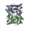
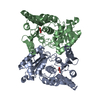
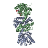
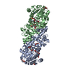
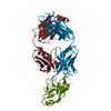
| Title | Structure of a transient complex between Dha-kinase subunits DhaM and DhaL from Lactococcus lactis | ||||||
 Components Components |
| ||||||
 Keywords Keywords |  TRANSFERASE / Transient Protein-protein complex TRANSFERASE complex PTS-DEPENDENT DIHYDROXYACETONE KINASE / ATP-binding / Glycerol metabolism / Nucleotide-binding / Phosphotransferase system TRANSFERASE / Transient Protein-protein complex TRANSFERASE complex PTS-DEPENDENT DIHYDROXYACETONE KINASE / ATP-binding / Glycerol metabolism / Nucleotide-binding / Phosphotransferase system | ||||||
| Function / homology |  Function and homology informationphosphoenolpyruvate-glycerone phosphotransferase / phosphoenolpyruvate-glycerone phosphotransferase activity / glycerone kinase activity / glycerol catabolic process / phosphoenolpyruvate-dependent sugar phosphotransferase system / phosphorylation / magnesium ion binding / ATP binding / membrane / cytoplasm Function and homology informationphosphoenolpyruvate-glycerone phosphotransferase / phosphoenolpyruvate-glycerone phosphotransferase activity / glycerone kinase activity / glycerol catabolic process / phosphoenolpyruvate-dependent sugar phosphotransferase system / phosphorylation / magnesium ion binding / ATP binding / membrane / cytoplasmSimilarity search - Function | ||||||
| Biological species |  Lactococcus lactis subsp. lactis (lactic acid bacteria) Lactococcus lactis subsp. lactis (lactic acid bacteria) | ||||||
| Method | X-RAY DIFFRACTION / SYNCHROTRON / MAD / Resolution: 2.1 Å | ||||||
 Authors Authors | Jeckelmann, J.M. / Zurbriggen, A. / Christen, S. / Baumann, U. / Erni, B. | ||||||
 Citation Citation | Journal: J.Biol.Chem. / Year: 2008 Title: X-ray Structures of the Three Lactococcus lactis Dihydroxyacetone Kinase Subunits and of a Transient Intersubunit Complex. Authors: Zurbriggen, A. / Jeckelmann, J.M. / Christen, S. / Bieniossek, C. / Baumann, U. / Erni, B. | ||||||
| History |
|
- Structure visualization
| Structure viewer | Molecule: MolmilJmol/JSmol |
|---|
- Downloads & links
-Download
| PDBx/mmCIF format | 3cr3.cif.gz | 130.2 KB | Display | PDBx/mmCIF format |
|---|---|---|---|---|
| PDB format | pdb3cr3.ent.gz | 108.4 KB | Display | PDB format |
| PDBx/mmJSON format | 3cr3.json.gz | Tree view | PDBx/mmJSON format | |
| Others |  Other downloads Other downloads |
-Validation report
| Arichive directory | https://data.pdbj.org/pub/pdb/validation_reports/cr/3cr3ftp://data.pdbj.org/pub/pdb/validation_reports/cr/3cr3 | HTTPS FTP |
|---|
-Related structure data


-Links
 PDBj
PDBj- Assembly
| Deposited unit | 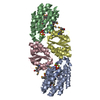
| ||||||||||||||||||||||||||||||||||||||||||||||||||||||||||
|---|---|---|---|---|---|---|---|---|---|---|---|---|---|---|---|---|---|---|---|---|---|---|---|---|---|---|---|---|---|---|---|---|---|---|---|---|---|---|---|---|---|---|---|---|---|---|---|---|---|---|---|---|---|---|---|---|---|---|---|
| 1 |
| ||||||||||||||||||||||||||||||||||||||||||||||||||||||||||
| 2 | 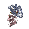
| ||||||||||||||||||||||||||||||||||||||||||||||||||||||||||
| 3 | 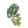
| ||||||||||||||||||||||||||||||||||||||||||||||||||||||||||
| 4 | 
| ||||||||||||||||||||||||||||||||||||||||||||||||||||||||||
| 5 | 
| ||||||||||||||||||||||||||||||||||||||||||||||||||||||||||
| Unit cell |
| ||||||||||||||||||||||||||||||||||||||||||||||||||||||||||
| Noncrystallographic symmetry (NCS) | NCS domain:
NCS domain segments: Component-ID: 1 / Refine code: 5
NCS ensembles :
|
-Components
| #1: Protein | Mass: 20909.639 Da / Num. of mol.: 2 Source method: isolated from a genetically manipulated source Details: lacI(q), oriR ColE1, bla Source: (gene. exp.) Lactococcus lactis subsp. lactis (lactic acid bacteria)Strain: IL1403 / Gene: dhaL / Plasmid: pMS-DhaKLM / Production host: Escherichia coli (E. coli) / Strain (production host): CB-delta-KLMReferences: UniProt: Q9CIV7, Transferases; Transferring phosphorus-containing groups#2: Protein | Mass: 13360.600 Da / Num. of mol.: 2 Source method: isolated from a genetically manipulated source Source: (gene. exp.) Lactococcus lactis subsp. lactis (lactic acid bacteria)Gene: dhaM / Plasmid: pMS-DhaKLM / Production host: Escherichia coli (E. coli) / Strain (production host): CB-delta-KLMReferences: UniProt: Q9CIV6, Transferases; Transferring phosphorus-containing groups; Phosphotransferases with an alcohol group as acceptor#3: Chemical | ChemComp-MG /   Mass: 24.305 Da / Num. of mol.: 4 / Source method: obtained synthetically / Formula: Mg Mass: 24.305 Da / Num. of mol.: 4 / Source method: obtained synthetically / Formula: Mg#4: Chemical | Adenosine diphosphate  Mass: 427.201 Da / Num. of mol.: 2 / Source method: obtained synthetically / Formula: C10H15N5O10P2 / Comment: ADP, energy-carrying molecule*YM Mass: 427.201 Da / Num. of mol.: 2 / Source method: obtained synthetically / Formula: C10H15N5O10P2 / Comment: ADP, energy-carrying molecule*YM#5: Water | ChemComp-HOH / | Water Mass: 18.015 Da / Num. of mol.: 192 / Source method: isolated from a natural source / Formula: H2O Mass: 18.015 Da / Num. of mol.: 192 / Source method: isolated from a natural source / Formula: H2O |
|---|
-Experimental details
-Experiment
| Experiment | Method: X-RAY DIFFRACTION / Number of used crystals: 1 |
|---|
- Sample preparation
| Crystal | Density Matthews: 2.32 Å3/Da / Density % sol: 47.03 % |
|---|---|
| Crystal grow | Temperature: 289 K / Method: vapor diffusion, sitting drop / pH: 8.5 Details: 0.1 M Tris/HCl, 0.2 M LiSO4, 36 % PEG 4000, pH 8.5, VAPOR DIFFUSION, SITTING DROP, temperature 289K |
-Data collection
| Diffraction | Mean temperature: 100 K | ||||||||||||
|---|---|---|---|---|---|---|---|---|---|---|---|---|---|
| Diffraction source | Source: SYNCHROTRON / Site: EMBL/DESY, Hamburg  / Beamline: X12 / Wavelength: 0.97885,0.97823,0.97240 / Beamline: X12 / Wavelength: 0.97885,0.97823,0.97240 | ||||||||||||
| Detector | Type: MARMOSAIC 225 mm CCD / Detector: CCD / Date: Dec 8, 2006 | ||||||||||||
| Radiation | Protocol: MAD / Monochromatic (M) / Laue (L): M / Scattering type: x-ray | ||||||||||||
| Radiation wavelength |
| ||||||||||||
| Reflection | Resolution: 2.1→147.442 Å / Num. all: 37982 / Num. obs: 37982 / % possible obs: 99.8 % / Observed criterion σ(F): 1 / Observed criterion σ(I): -3 / Redundancy: 4.9 % / Biso Wilson estimate: 26.5 Å2 / Rmerge(I) obs: 0.047 / Net I/σ(I): 22.2 | ||||||||||||
| Reflection shell | Resolution: 2.1→2.21 Å / % possible obs: 100 % / Redundancy: 4.1 % / Rmerge(I) obs: 0.161 / Mean I/σ(I) obs: 8.1 / Num. unique all: 5440 / Rsym value: 0.214 / % possible all: 24.4 |
- Processing
| Software |
| |||||||||||||||||||||||||||||||||||||||||||||||||||||||||||||||||||||||||||||||||||||||||||||||
|---|---|---|---|---|---|---|---|---|---|---|---|---|---|---|---|---|---|---|---|---|---|---|---|---|---|---|---|---|---|---|---|---|---|---|---|---|---|---|---|---|---|---|---|---|---|---|---|---|---|---|---|---|---|---|---|---|---|---|---|---|---|---|---|---|---|---|---|---|---|---|---|---|---|---|---|---|---|---|---|---|---|---|---|---|---|---|---|---|---|---|---|---|---|---|---|---|
| Refinement | Method to determine structure: MAD / Resolution: 2.1→66.23 Å / Cor.coef. Fo:Fc: 0.929 / Cor.coef. Fo:Fc free: 0.898 / SU B: 9.876 / SU ML: 0.144 / Cross valid method: THROUGHOUT / σ(F): 0 / ESU R: 0.261 / ESU R Free: 0.209 / Stereochemistry target values: MAXIMUM LIKELIHOOD
| |||||||||||||||||||||||||||||||||||||||||||||||||||||||||||||||||||||||||||||||||||||||||||||||
| Solvent computation | Ion probe radii: 0.8 Å / Shrinkage radii: 0.8 Å / VDW probe radii: 1.4 Å / Solvent model: MASK | |||||||||||||||||||||||||||||||||||||||||||||||||||||||||||||||||||||||||||||||||||||||||||||||
| Displacement parameters | Biso mean: 19.17 Å2
| |||||||||||||||||||||||||||||||||||||||||||||||||||||||||||||||||||||||||||||||||||||||||||||||
| Refinement step | Cycle: LAST / Resolution: 2.1→66.23 Å
| |||||||||||||||||||||||||||||||||||||||||||||||||||||||||||||||||||||||||||||||||||||||||||||||
| Refine LS restraints |
| |||||||||||||||||||||||||||||||||||||||||||||||||||||||||||||||||||||||||||||||||||||||||||||||
| Refine LS restraints NCS | Dom-ID: 1 / Refine-ID: X-RAY DIFFRACTION
| |||||||||||||||||||||||||||||||||||||||||||||||||||||||||||||||||||||||||||||||||||||||||||||||
| LS refinement shell | Resolution: 2.1→2.155 Å / Total num. of bins used: 20
|
