Mass: 18.015 Da / Num. of mol.: 2 / Source method: isolated from a natural source / Formula: H2O
-
Experimental details
-
Experiment
Experiment
Method: X-RAY DIFFRACTION / Number of used crystals: 1
-
Sample preparation
Crystal
Density Matthews: 3.8 Å3/Da / Density % sol: 67.65 %
Crystal grow
Temperature: 298 K / Method: vapor diffusion, hanging drop / pH: 6.5 Details: 1.5 M (NH4)SO4, 0.1 M Bis-Tris, 0.1 M NaCl, pH 6.5, VAPOR DIFFUSION, HANGING DROP, temperature 298K
-
Data collection
Diffraction
Mean temperature: 100 K
Diffraction source
Source: SYNCHROTRON / Site: ALS / Beamline: 8.2.2 / Wavelength: 1 Å
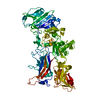
Method to determine structure: MOLECULAR REPLACEMENT / Resolution: 3.9→74.33 Å / Cor.coef. Fo:Fc: 0.897 / Cor.coef. Fo:Fc free: 0.86 / SU B: 93.365 / SU ML: 0.617 / TLS residual ADP flag: LIKELY RESIDUAL / Cross valid method: THROUGHOUT / σ(F): 0 / ESU R Free: 1.083 / Stereochemistry target values: MAXIMUM LIKELIHOOD Details: A peptide cross-link is formed by the toxin between Lys50 and Glu270 of neighboring actin molecules. The cross-link is not modeled in the coordinate set due to disorder. HYDROGENS HAVE BEEN ...Details: A peptide cross-link is formed by the toxin between Lys50 and Glu270 of neighboring actin molecules. The cross-link is not modeled in the coordinate set due to disorder. HYDROGENS HAVE BEEN ADDED IN THE RIDING POSITIONS. Ellipsoidal truncation and anisotropic scale factors have been applied to the structure factors and used in refinement. The ellipsoid has principle axes of 5.8, 3.9, and 3.9 reciprocal angstroms along a*, b*, and c*, respectively. The submitted structure factor archive contains the truncated/scaled structure factors and the original, unmodified intensities.
Rfactor
Num. reflection
% reflection
Selection details
Rfree
0.278
400
4.7 %
RANDOM
Rwork
0.223
-
-
-
obs
0.226
8450
68.26 %
-
Solvent computation
Ion probe radii: 0.8 Å / Shrinkage radii: 0.8 Å / VDW probe radii: 1.4 Å / Solvent model: MASK
Displacement parameters
Biso mean: 47.869 Å2
Baniso -1
Baniso -2
Baniso -3
1-
-13.77 Å2
0 Å2
0 Å2
2-
-
6.73 Å2
0 Å2
3-
-
-
7.04 Å2
Refine analyze
Luzzati coordinate error free: 1.083 Å
Refinement step
Cycle: LAST / Resolution: 3.9→74.33 Å
Protein
Nucleic acid
Ligand
Solvent
Total
Num. atoms
5936
0
91
2
6029
Refine LS restraints
Refine-ID
Type
Dev ideal
Dev ideal target
Number
X-RAY DIFFRACTION
r_bond_refined_d
0.006
0.022
6159
X-RAY DIFFRACTION
r_bond_other_d
0.007
0.02
4082
X-RAY DIFFRACTION
r_angle_refined_deg
0.928
1.969
8372
X-RAY DIFFRACTION
r_angle_other_deg
0.78
3.001
9931
X-RAY DIFFRACTION
r_dihedral_angle_1_deg
2.853
5
751
X-RAY DIFFRACTION
r_dihedral_angle_2_deg
26.615
24.143
280
X-RAY DIFFRACTION
r_dihedral_angle_3_deg
11.866
15
1007
X-RAY DIFFRACTION
r_dihedral_angle_4_deg
9.353
15
34
X-RAY DIFFRACTION
r_chiral_restr
0.054
0.2
928
X-RAY DIFFRACTION
r_gen_planes_refined
0.003
0.021
6808
X-RAY DIFFRACTION
r_gen_planes_other
0.001
0.02
1252
X-RAY DIFFRACTION
r_mcbond_it
1.103
2
3753
X-RAY DIFFRACTION
r_mcbond_other
0.05
2
1526
X-RAY DIFFRACTION
r_mcangle_it
1.587
3
6063
X-RAY DIFFRACTION
r_scbond_it
0.323
2
2406
X-RAY DIFFRACTION
r_scangle_it
0.567
3
2309
LS refinement shell
Resolution: 3.9→3.998 Å / Total num. of bins used: 20
Rfactor
Num. reflection
% reflection
Rfree
0.284
10
-
Rwork
0.263
137
-
all
-
147
-
obs
-
-
16.63 %
Refinement TLS params.
Method: refined / Refine-ID: X-RAY DIFFRACTION
ID
L11 (°2)
L12 (°2)
L13 (°2)
L22 (°2)
L23 (°2)
L33 (°2)
S11 (Å °)
S12 (Å °)
S13 (Å °)
S21 (Å °)
S22 (Å °)
S23 (Å °)
S31 (Å °)
S32 (Å °)
S33 (Å °)
T11 (Å2)
T12 (Å2)
T13 (Å2)
T22 (Å2)
T23 (Å2)
T33 (Å2)
Origin x (Å)
Origin y (Å)
Origin z (Å)
1
0.5605
-0.3393
0.1322
1.0272
0.1281
0.4917
0.039
0.0479
0.047
0.0318
-0.0318
-0.1274
0.0521
0.0355
-0.0071
0.1882
0.0021
-0.0018
-0.1606
0.0396
-0.2401
-4.905
37.904
-5.469
2
3.7029
-0.3763
-0.3273
0.8785
-0.116
3.0652
0.0348
0.1458
0.0265
-0.0355
-0.0346
0.0663
-0.0287
-0.1009
-0.0002
0.1538
-0.0265
-0.0135
-0.3667
-0.0538
-0.2682
6.7754
-3.7914
-19.2784
3
3.475
0.1081
0.6509
1.5101
0.6517
0.5588
0.0964
-0.1666
0.4189
-0.1374
0.3237
-0.8476
-0.4237
0.3738
-0.4201
0.1995
-0.0835
0.1071
-0.3065
0.0051
0.0184
8.0319
67.792
-3.3341
Refinement TLS group
ID
Refine-ID
Refine TLS-ID
Auth asym-ID
Label asym-ID
Auth seq-ID
Label seq-ID
1
X-RAY DIFFRACTION
1
A
A
1 - 372
3 - 374
2
X-RAY DIFFRACTION
2
D
B
1 - 260
1 - 260
3
X-RAY DIFFRACTION
3
G
C
1 - 125
1 - 125
+
About Yorodumi
-
News
-
Feb 9, 2022. New format data for meta-information of EMDB entries
New format data for meta-information of EMDB entries
Version 3 of the EMDB header file is now the official format.
The previous official version 1.9 will be removed from the archive.
In the structure databanks used in Yorodumi, some data are registered as the other names, "COVID-19 virus" and "2019-nCoV". Here are the details of the virus and the list of structure data.
Jan 31, 2019. EMDB accession codes are about to change! (news from PDBe EMDB page)
EMDB accession codes are about to change! (news from PDBe EMDB page)
The allocation of 4 digits for EMDB accession codes will soon come to an end. Whilst these codes will remain in use, new EMDB accession codes will include an additional digit and will expand incrementally as the available range of codes is exhausted. The current 4-digit format prefixed with “EMD-” (i.e. EMD-XXXX) will advance to a 5-digit format (i.e. EMD-XXXXX), and so on. It is currently estimated that the 4-digit codes will be depleted around Spring 2019, at which point the 5-digit format will come into force.
The EM Navigator/Yorodumi systems omit the EMD- prefix.
Related info.:Q: What is EMD? / ID/Accession-code notation in Yorodumi/EM Navigator
Yorodumi is a browser for structure data from EMDB, PDB, SASBDB, etc.
This page is also the successor to EM Navigator detail page, and also detail information page/front-end page for Omokage search.
The word "yorodu" (or yorozu) is an old Japanese word meaning "ten thousand". "mi" (miru) is to see.
Related info.:EMDB / PDB / SASBDB / Comparison of 3 databanks / Yorodumi Search / Aug 31, 2016. New EM Navigator & Yorodumi / Yorodumi Papers / Jmol/JSmol / Function and homology information / Changes in new EM Navigator and Yorodumi
 Movie
Movie Controller

 Yorodumi
 Open data
 Basic information
 Components
 Keywords
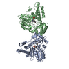
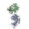
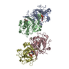
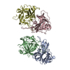
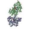
 Structural protein/Hydrolase / cross-linked dimer / ATP-binding /
  Function and homology information

 Authors
 Citation
 Structure visualization
 Downloads & links
 Other downloads


 PDBj
PDBj


 Assembly
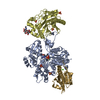


 Mass: 40.078 Da / Num. of mol.: 2 / Source method: obtained synthetically / Formula: Ca
 Mass: 96.063 Da / Num. of mol.: 6 / Source method: obtained synthetically / Formula: SO4
 Mass: 507.181 Da / Num. of mol.: 1 / Source method: obtained synthetically / Formula: C10H16N5O13P3 / Comment: ATP, energy-carrying molecule*YM
 Sample preparation
 / Beamline: 8.2.2 / Wavelength: 1 Å
 Processing