 Movie
Movie Controller

[English] 日本語
 Yorodumi
- PDB-3c7i: X-RAY crystal structure of the complex between the grb2-sh2 domai... -
+ Open data

- Basic information
| Entry | Database: PDB / ID: 3c7i | |||||||||
|---|---|---|---|---|---|---|---|---|---|---|
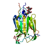
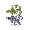
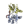
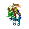
| Title | X-RAY crystal structure of the complex between the grb2-sh2 domain and a flexible ligand, FPTVN. | |||||||||
 Components Components | Growth factor receptor-bound protein 2 GRB2 GRB2 | |||||||||
 Keywords Keywords | SPLICING / FLEXIBLE / CONSTRAINED / ENTROPY / GRB2-SH2 / LIGAND PREORGANIZATION / Alternative splicing / Host-virus interaction / Phosphoprotein / SH2 domain / SH3 domain | |||||||||
| Function / homology |  Function and homology information Function and homology informationguanyl-nucleotide exchange factor adaptor activity / Grb2-EGFR complex / branching involved in labyrinthine layer morphogenesis / STAT5 Activation / COP9 signalosome / vesicle membrane / neurotrophin TRKA receptor binding / Activated NTRK2 signals through PI3K / MET receptor recycling / transmembrane receptor protein tyrosine kinase adaptor activity ...guanyl-nucleotide exchange factor adaptor activity / Grb2-EGFR complex / branching involved in labyrinthine layer morphogenesis / STAT5 Activation / COP9 signalosome / vesicle membrane / neurotrophin TRKA receptor binding / Activated NTRK2 signals through PI3K / MET receptor recycling / transmembrane receptor protein tyrosine kinase adaptor activity / Signaling by cytosolic FGFR1 fusion mutants / Interleukin-15 signaling / MET activates PTPN11 / MET activates RAP1 and RAC1 / CD28 dependent Vav1 pathway / Costimulation by the CD28 family / MET activates PI3K/AKT signaling / Signal regulatory protein family interactions / epidermal growth factor receptor binding / Regulation of KIT signaling / positive regulation of actin filament polymerization / PI-3K cascade:FGFR3 / STAT5 activation downstream of FLT3 ITD mutants / PI-3K cascade:FGFR2 / PI-3K cascade:FGFR4 / PI-3K cascade:FGFR1 / endodermal cell differentiation / regulation of MAPK cascade / GRB2:SOS provides linkage to MAPK signaling for Integrins / RHOU GTPase cycle / PI3K events in ERBB2 signaling / Signaling by ALK fusions and activated point mutants / SOS-mediated signalling / Activated NTRK3 signals through RAS / RET signaling / Activated NTRK2 signals through RAS / insulin receptor substrate binding / PI3K Cascade / Interleukin-3, Interleukin-5 and GM-CSF signaling / SHC1 events in ERBB4 signaling / RHO GTPases Activate WASPs and WAVEs / Signalling to RAS / fibroblast growth factor receptor signaling pathway / GAB1 signalosome / SHC-related events triggered by IGF1R / Activated NTRK2 signals through FRS2 and FRS3 / Role of LAT2/NTAL/LAB on calcium mobilization / Signal attenuation / Interleukin receptor SHC signaling / SHC-mediated cascade:FGFR3 / MET activates RAS signaling / Schwann cell development / Signaling by PDGFRA transmembrane, juxtamembrane and kinase domain mutants / Signaling by PDGFRA extracellular domain mutants / SHC-mediated cascade:FGFR2 / SHC-mediated cascade:FGFR4 / Signaling by FGFR4 in disease / SHC-mediated cascade:FGFR1 / Erythropoietin activates RAS / signal transduction in response to DNA damage / FRS-mediated FGFR3 signaling / Signaling by CSF3 (G-CSF) / Signaling by FLT3 ITD and TKD mutants / FRS-mediated FGFR2 signaling / FRS-mediated FGFR4 signaling / Signaling by FGFR3 in disease / FRS-mediated FGFR1 signaling / Tie2 Signaling / Signaling by FGFR2 in disease / GRB2 events in EGFR signaling / SHC1 events in EGFR signaling / myelination / EGFR Transactivation by Gastrin / Signaling by FLT3 fusion proteins / FLT3 Signaling / Signaling by FGFR1 in disease / GRB2 events in ERBB2 signaling / NCAM signaling for neurite out-growth / phosphotyrosine residue binding / ephrin receptor binding / SHC1 events in ERBB2 signaling / Downstream signal transduction / Insulin receptor signalling cascade / Constitutive Signaling by Overexpressed ERBB2 / FCERI mediated Ca+2 mobilization / InlB-mediated entry of Listeria monocytogenes into host cell / insulin-like growth factor receptor signaling pathway / Signaling by phosphorylated juxtamembrane, extracellular and kinase domain KIT mutants / Antigen activates B Cell Receptor (BCR) leading to generation of second messengers / cellular response to ionizing radiation / Regulation of signaling by CBL / Negative regulation of FGFR3 signaling / FCGR3A-mediated phagocytosis / FCERI mediated MAPK activation / Negative regulation of FGFR2 signaling / Negative regulation of FGFR4 signaling / EGFR downregulation / Negative regulation of FGFR1 signaling / B cell receptor signaling pathway / Signaling by ERBB2 TMD/JMD mutantsSimilarity search - Function | |||||||||
| Biological species |  Homo sapiens (human) Homo sapiens (human) | |||||||||
| Method | X-RAY DIFFRACTION / MOLECULAR REPLACEMENT / Resolution: 1.7 Å | |||||||||
 Authors Authors | Benfield, A.P. / Martin, S.F. | |||||||||
 Citation Citation | Journal: Angew.Chem.Int.Ed.Engl. / Year: 2006 Title: Ligand Preorganization May be Accompanied by Entropic Penalties in Protein-Ligand Interactions. Authors: Benfield, A.P. / Teresk, M.G. / Plake, H.R. / Delorbe, J.E. / Millspaugh, L.E. / Martin, S.F. | |||||||||
| History |
|
- Structure visualization
| Structure viewer | Molecule: MolmilJmol/JSmol |
|---|
- Downloads & links
-Download
| PDBx/mmCIF format | 3c7i.cif.gz | 38.3 KB | Display | PDBx/mmCIF format |
|---|---|---|---|---|
| PDB format | pdb3c7i.ent.gz | 25.1 KB | Display | PDB format |
| PDBx/mmJSON format | 3c7i.json.gz | Tree view | PDBx/mmJSON format | |
| Others |  Other downloads Other downloads |
-Validation report
| Arichive directory | https://data.pdbj.org/pub/pdb/validation_reports/c7/3c7iftp://data.pdbj.org/pub/pdb/validation_reports/c7/3c7i | HTTPS FTP |
|---|
-Related structure data


-Links
 PDBj
PDBj


- Assembly
| Deposited unit | 
| ||||||||
|---|---|---|---|---|---|---|---|---|---|
| 1 | 
| ||||||||
| Unit cell |
|
-Components
| #1: Protein | GRB2 / Adapter protein GRB2 / SH2/SH3 adapter GRB2 / Protein Ash Mass: 13687.465 Da / Num. of mol.: 1 Source method: isolated from a genetically manipulated source Source: (gene. exp.) Homo sapiens (human) / Gene: GRB2, ASH / Production host:  ESCHERICHIA COLI (E. coli) / References: UniProt: P62993 ESCHERICHIA COLI (E. coli) / References: UniProt: P62993 |
|---|---|
| #2: Chemical | ChemComp-TVN /   Mass: 529.481 Da / Num. of mol.: 1 / Source method: obtained synthetically / Formula: C21H32N5O9P Mass: 529.481 Da / Num. of mol.: 1 / Source method: obtained synthetically / Formula: C21H32N5O9P |
| #3: Chemical | ChemComp-FMT / Formic acid  Mass: 46.025 Da / Num. of mol.: 1 / Source method: obtained synthetically / Formula: CH2O2 Mass: 46.025 Da / Num. of mol.: 1 / Source method: obtained synthetically / Formula: CH2O2 |
| #4: Water | ChemComp-HOH / Water Mass: 18.015 Da / Num. of mol.: 121 / Source method: isolated from a natural source / Formula: H2O Mass: 18.015 Da / Num. of mol.: 121 / Source method: isolated from a natural source / Formula: H2O |
-Experimental details
-Experiment
| Experiment | Method: X-RAY DIFFRACTION / Number of used crystals: 1 |
|---|
- Sample preparation
| Crystal | Density Matthews: 1.77 Å3/Da / Density % sol: 30.4 % |
|---|---|
| Crystal grow | Temperature: 293 K / Method: vapor diffusion, hanging drop / pH: 5 Details: LIGAND (FPTVN) was added in a 1.5 molar excess to 15 MG/ML GRB2-SH2 in pure water. This solution was mixed with an equal volume of 4.0 m sodium formate., pH 5.00, VAPOR DIFFUSION, HANGING DROP, temperature 293KK |
-Data collection
| Diffraction | Mean temperature: 100 K |
|---|---|
| Diffraction source | Source: ROTATING ANODE / Type: RIGAKU RU200 / Wavelength: 1.5418 |
| Detector | Type: RIGAKU RAXIS IV / Detector: IMAGE PLATE / Date: Jan 1, 2005 |
| Radiation | Protocol: SINGLE WAVELENGTH / Monochromatic (M) / Laue (L): M / Scattering type: x-ray |
| Radiation wavelength | Wavelength: 1.5418 Å / Relative weight: 1 |
| Reflection | Resolution: 1.7→30 Å / Num. obs: 11224 / % possible obs: 97.2 % / Observed criterion σ(I): 1 / Redundancy: 5.2 % / Rmerge(I) obs: 0.054 / Net I/σ(I): 23.3 |
| Reflection shell | Resolution: 1.7→1.76 Å / Redundancy: 2.6 % / Rmerge(I) obs: 0.174 / % possible all: 79.7 |
- Processing
| Software |
| ||||||||||||||||||||||||||||||||||||||||||||||||||||||||||||||||||||||||||||||||
|---|---|---|---|---|---|---|---|---|---|---|---|---|---|---|---|---|---|---|---|---|---|---|---|---|---|---|---|---|---|---|---|---|---|---|---|---|---|---|---|---|---|---|---|---|---|---|---|---|---|---|---|---|---|---|---|---|---|---|---|---|---|---|---|---|---|---|---|---|---|---|---|---|---|---|---|---|---|---|---|---|---|
| Refinement | Method to determine structure: MOLECULAR REPLACEMENT / Resolution: 1.7→14.7 Å / σ(F): 0 / Stereochemistry target values: ENGH & HUBER
| ||||||||||||||||||||||||||||||||||||||||||||||||||||||||||||||||||||||||||||||||
| Solvent computation | Bsol: 49.5 Å2 | ||||||||||||||||||||||||||||||||||||||||||||||||||||||||||||||||||||||||||||||||
| Displacement parameters | Biso mean: 20.68 Å2
| ||||||||||||||||||||||||||||||||||||||||||||||||||||||||||||||||||||||||||||||||
| Refinement step | Cycle: LAST / Resolution: 1.7→14.7 Å
| ||||||||||||||||||||||||||||||||||||||||||||||||||||||||||||||||||||||||||||||||
| Refine LS restraints |
| ||||||||||||||||||||||||||||||||||||||||||||||||||||||||||||||||||||||||||||||||
| LS refinement shell | Resolution: 1.7→1.75 Å / Total num. of bins used: 11 /
| ||||||||||||||||||||||||||||||||||||||||||||||||||||||||||||||||||||||||||||||||
| Xplor file |
|
