 Movie
Movie Controller


+ Open data

- Basic information
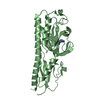
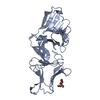
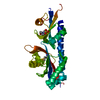
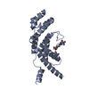
| Entry | Database: PDB / ID: 3bmp | |||||||||
|---|---|---|---|---|---|---|---|---|---|---|
| Title | HUMAN BONE MORPHOGENETIC PROTEIN-2 (BMP-2) | |||||||||
 Components Components | PROTEIN (BONE MORPHOGENETIC PROTEIN 2 (BMP-2)) | |||||||||
 Keywords Keywords |  CYTOKINE / BONE MORPHOGENETIC PROTEIN / CYSTIN-KNOT / TGFB-FAMILY CYTOKINE / BONE MORPHOGENETIC PROTEIN / CYSTIN-KNOT / TGFB-FAMILY | |||||||||
| Function / homology |  Function and homology information Function and homology informationcardiac atrium formation / cardiocyte differentiation / negative regulation of calcium-independent cell-cell adhesion / cardiac jelly development / negative regulation of aldosterone biosynthetic process / embryonic heart tube anterior/posterior pattern specification / atrioventricular canal morphogenesis / negative regulation of cortisol biosynthetic process / mesenchymal cell proliferation involved in ureteric bud development / negative regulation of steroid biosynthetic process ...cardiac atrium formation / cardiocyte differentiation / negative regulation of calcium-independent cell-cell adhesion / cardiac jelly development / negative regulation of aldosterone biosynthetic process / embryonic heart tube anterior/posterior pattern specification / atrioventricular canal morphogenesis / negative regulation of cortisol biosynthetic process / mesenchymal cell proliferation involved in ureteric bud development / negative regulation of steroid biosynthetic process / ameloblast differentiation / positive regulation of extracellular matrix constituent secretion / negative regulation of cardiac muscle cell differentiation / positive regulation of odontogenesis / regulation of odontogenesis of dentin-containing tooth / endodermal-mesodermal cell signaling / corticotropin hormone secreting cell differentiation / lung vasculature development / negative regulation of insulin-like growth factor receptor signaling pathway / mesenchyme development / thyroid-stimulating hormone-secreting cell differentiation / aortic valve development / telencephalon regionalization / positive regulation of phosphatase activity / positive regulation of cartilage development / proteoglycan metabolic process / heart induction / positive regulation of peroxisome proliferator activated receptor signaling pathway / pericardium development / BMP receptor complex / co-receptor binding / telencephalon development / cardiac epithelial to mesenchymal transition / BMP receptor binding / positive regulation of odontoblast differentiation / mesenchymal cell differentiation / endocardial cushion formation / positive regulation of bone mineralization involved in bone maturation / Transcriptional regulation by RUNX2 / phosphatase activator activity / positive regulation of astrocyte differentiation / Signaling by BMP / cardiac muscle cell differentiation / cellular response to BMP stimulus / cardiac muscle tissue morphogenesis / positive regulation of ossification / astrocyte differentiation / Molecules associated with elastic fibres / atrioventricular valve morphogenesis / positive regulation of p38MAPK cascade / endocardial cushion morphogenesis / negative regulation of fat cell differentiation / branching involved in ureteric bud morphogenesis / bone mineralization / odontogenesis of dentin-containing tooth / positive regulation of osteoblast proliferation / inner ear development / positive regulation of SMAD protein signal transduction / positive regulation of Wnt signaling pathway / cellular response to organic cyclic compound / negative regulation of cell cycle / epithelial to mesenchymal transition / positive regulation of epithelial to mesenchymal transition / positive regulation of fat cell differentiation / positive regulation of osteoblast differentiation / cell fate commitment / chondrocyte differentiation / BMP signaling pathway / positive regulation of bone mineralization / Notch signaling pathway / positive regulation of neuron differentiation / protein serine/threonine kinase activator activity / osteoclast differentiation / negative regulation of MAP kinase activity / skeletal system development / cytokine activity / negative regulation of smooth muscle cell proliferation / response to bacterium / animal organ morphogenesis / negative regulation of transforming growth factor beta receptor signaling pathway / negative regulation of canonical Wnt signaling pathway / growth factor activity / protein destabilization / bone development / osteoblast differentiation / positive regulation of miRNA transcription / Regulation of RUNX2 expression and activity / positive regulation of DNA-binding transcription factor activity / cell-cell signaling / heart development / positive regulation of protein binding / in utero embryonic development / positive regulation of MAPK cascade / transcription by RNA polymerase II / positive regulation of ERK1 and ERK2 cascade / response to hypoxia / positive regulation of cell migration / inflammatory response / positive regulation of protein phosphorylation / positive regulation of apoptotic processSimilarity search - Function | |||||||||
| Biological species |  Homo sapiens (human) Homo sapiens (human) | |||||||||
| Method | X-RAY DIFFRACTION / MOLECULAR REPLACEMENT / Resolution: 2.7 Å | |||||||||
 Authors Authors | Scheufler, C. / Sebald, W. / Huelsmeyer, M. | |||||||||
 Citation Citation | Journal: J.Mol.Biol. / Year: 1999 Title: Crystal structure of human bone morphogenetic protein-2 at 2.7 A resolution. Authors: Scheufler, C. / Sebald, W. / Hulsmeyer, M. #1: Journal: Eur.J.Biochem. / Year: 1996Title: Human Bone Morphogenetic Protein 2 Contains a Heparin-Binding Site which Modifies its Biological Activity Authors: Ruppert, R. / Hoffmann, E. / Sebald, W. | |||||||||
| History |
|
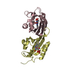
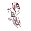
- Structure visualization
| Structure viewer | Molecule: MolmilJmol/JSmol |
|---|
- Downloads & links
-Download
| PDBx/mmCIF format | 3bmp.cif.gz | 35.1 KB | Display | PDBx/mmCIF format |
|---|---|---|---|---|
| PDB format | pdb3bmp.ent.gz | 23.3 KB | Display | PDB format |
| PDBx/mmJSON format | 3bmp.json.gz | Tree view | PDBx/mmJSON format | |
| Others |  Other downloads Other downloads |
-Validation report
| Arichive directory | https://data.pdbj.org/pub/pdb/validation_reports/bm/3bmpftp://data.pdbj.org/pub/pdb/validation_reports/bm/3bmp | HTTPS FTP |
|---|
-Related structure data
| Related structure data |  1tfgS S: Starting model for refinement |
|---|---|
| Similar structure data |


-Links
 PDBj
PDBj


- Assembly
| Deposited unit | 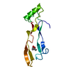
| ||||||||
|---|---|---|---|---|---|---|---|---|---|
| 1 | 
| ||||||||
| Unit cell |
| ||||||||
| Details | THE NATIVE HOMODIMERIC FORM OF BMP-2 IS BUILT BY CRYSTALLOGRAPHIC SYMMETRY. THE MONOMERS ARE LINKED VIA A CYSTINE BRIDGE (CYS78 FROM BOTH SUBUNITS) |
-Components
| #1: Protein | Mass: 12923.854 Da / Num. of mol.: 1 Source method: isolated from a genetically manipulated source Source: (gene. exp.) Homo sapiens (human) / Production host:  Escherichia coli (E. coli) / References: UniProt: P12643 Escherichia coli (E. coli) / References: UniProt: P12643 |
|---|---|
| #2: Chemical | ChemComp-MPD / (2-Methyl-2,4-pentanediol  Mass: 118.174 Da / Num. of mol.: 1 / Source method: obtained synthetically / Formula: C6H14O2 / Comment: precipitant*YM Mass: 118.174 Da / Num. of mol.: 1 / Source method: obtained synthetically / Formula: C6H14O2 / Comment: precipitant*YM |
| #3: Water | ChemComp-HOH / Water Mass: 18.015 Da / Num. of mol.: 33 / Source method: isolated from a natural source / Formula: H2O Mass: 18.015 Da / Num. of mol.: 33 / Source method: isolated from a natural source / Formula: H2O |
-Experimental details
-Experiment
| Experiment | Method: X-RAY DIFFRACTION / Number of used crystals: 1 |
|---|
- Sample preparation
| Crystal | Density Matthews: 3 Å3/Da / Density % sol: 59 % | ||||||||||||||||||||||||||||||
|---|---|---|---|---|---|---|---|---|---|---|---|---|---|---|---|---|---|---|---|---|---|---|---|---|---|---|---|---|---|---|---|
| Crystal grow | pH: 5.4 Details: CRYSTALLIZATION CONDITIONS: PROTEIN WAS CRYSTALLIZED FROM 100 MM LITHIUM SULFATE, 12% TERT-BUTANOL, 50 MM CITRATE, PH 5.4 | ||||||||||||||||||||||||||||||
| Crystal grow | *PLUS Temperature: 20 ℃ / Method: vapor diffusion, hanging drop | ||||||||||||||||||||||||||||||
| Components of the solutions | *PLUS
|
-Data collection
| Diffraction | Mean temperature: 100 K |
|---|---|
| Diffraction source | Source: ROTATING ANODE / Type: RIGAKU RU200 / Wavelength: 1.5418 |
| Detector | Type: SIEMENS / Detector: AREA DETECTOR / Date: Nov 30, 1997 / Details: COLLIMATOR |
| Radiation | Monochromator: GRAPHITE / Protocol: SINGLE WAVELENGTH / Monochromatic (M) / Laue (L): M / Scattering type: x-ray |
| Radiation wavelength | Wavelength: 1.5418 Å / Relative weight: 1 |
| Reflection | Resolution: 2.7→10 Å / Num. obs: 4647 / % possible obs: 94.6 % / Observed criterion σ(I): 0 / Redundancy: 3 % / Biso Wilson estimate: 60.3 Å2 / Rmerge(I) obs: 0.041 / Net I/σ(I): 24.2 |
| Reflection shell | Resolution: 2.7→2.8 Å / Redundancy: 2.2 % / Rmerge(I) obs: 0.123 / Mean I/σ(I) obs: 5.4 / % possible all: 82.2 |
| Reflection shell | *PLUS % possible obs: 82.2 % |
- Processing
| Software |
| ||||||||||||||||||||||||||||||||||||||||||||||||||||||||||||||||||||||||||||||||
|---|---|---|---|---|---|---|---|---|---|---|---|---|---|---|---|---|---|---|---|---|---|---|---|---|---|---|---|---|---|---|---|---|---|---|---|---|---|---|---|---|---|---|---|---|---|---|---|---|---|---|---|---|---|---|---|---|---|---|---|---|---|---|---|---|---|---|---|---|---|---|---|---|---|---|---|---|---|---|---|---|---|
| Refinement | Method to determine structure: MOLECULAR REPLACEMENT Starting model: 1TFG Resolution: 2.7→25 Å / Rfactor Rfree error: 0.016 / Cross valid method: THROUGHOUT / σ(F): 0
| ||||||||||||||||||||||||||||||||||||||||||||||||||||||||||||||||||||||||||||||||
| Solvent computation | Solvent model: FLAT MODEL / Bsol: 44.6 Å2 / ksol: 0.35 e/Å3 | ||||||||||||||||||||||||||||||||||||||||||||||||||||||||||||||||||||||||||||||||
| Displacement parameters | Biso mean: 37 Å2
| ||||||||||||||||||||||||||||||||||||||||||||||||||||||||||||||||||||||||||||||||
| Refine analyze |
| ||||||||||||||||||||||||||||||||||||||||||||||||||||||||||||||||||||||||||||||||
| Refinement step | Cycle: LAST / Resolution: 2.7→25 Å
| ||||||||||||||||||||||||||||||||||||||||||||||||||||||||||||||||||||||||||||||||
| Refine LS restraints |
| ||||||||||||||||||||||||||||||||||||||||||||||||||||||||||||||||||||||||||||||||
| LS refinement shell | Resolution: 2.7→2.87 Å / Rfactor Rfree error: 0.064 / Total num. of bins used: 6
| ||||||||||||||||||||||||||||||||||||||||||||||||||||||||||||||||||||||||||||||||
| Software | *PLUS Name: CNS / Version: 0.5 / Classification: refinement | ||||||||||||||||||||||||||||||||||||||||||||||||||||||||||||||||||||||||||||||||
| Refine LS restraints | *PLUS
|
