DNA (5'-D(*DTP*DTP*DTP*DTP*DAP*DTP*DAP*DCP*DGP*DAP*DTP*DGP*DGP*DG)-3')
DNA (5'-D(P*DCP*DCP*DCP*DAP*DTP*DCP*DGP*DTP*DAP*DT)-3')
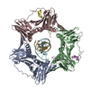
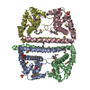
DNA polymerase III subunit betaDNA polymerase III holoenzyme
Keywords
TRANSFERASE / TRANSCRIPTION/DNA / beta subunit / sliding clamp / E. coli polymerase III / DNA complex / TRANSCRIPTION-DNA COMPLEX
Function / homology
Function and homology information
Hda-beta clamp complex / bacterial-type DNA replication / replication inhibiting complex / DNA polymerase III complex / replisome / regulation of DNA-templated DNA replication initiation / DNA strand elongation involved in DNA replication / error-prone translesion synthesis / negative regulation of DNA-templated DNA replication initiation / 3'-5' exonuclease activity ...Hda-beta clamp complex / bacterial-type DNA replication / replication inhibiting complex / DNA polymerase III complex / replisome / regulation of DNA-templated DNA replication initiation / DNA strand elongation involved in DNA replication / error-prone translesion synthesis / negative regulation of DNA-templated DNA replication initiation / 3'-5' exonuclease activity / DNA-templated DNA replication / DNA-directed DNA polymerase activity / DNA damage response / protein homodimerization activity / DNA binding / identical protein binding / cytosol Similarity search - Function
DNA Polymerase III; Chain A, domain 2 / DNA Polymerase III, subunit A, domain 2 / DNA polymerase III, beta sliding clamp / DNA polymerase III, beta sliding clamp, N-terminal / DNA polymerase III, beta sliding clamp, C-terminal / DNA polymerase III, beta sliding clamp, central / DNA polymerase III beta subunit, N-terminal domain / DNA polymerase III beta subunit, central domain / DNA polymerase III beta subunit, C-terminal domain / DNA polymerase III beta subunit ...DNA Polymerase III; Chain A, domain 2 / DNA Polymerase III, subunit A, domain 2 / DNA polymerase III, beta sliding clamp / DNA polymerase III, beta sliding clamp, N-terminal / DNA polymerase III, beta sliding clamp, C-terminal / DNA polymerase III, beta sliding clamp, central / DNA polymerase III beta subunit, N-terminal domain / DNA polymerase III beta subunit, central domain / DNA polymerase III beta subunit, C-terminal domain / DNA polymerase III beta subunit / : / Roll / Alpha Beta Similarity search - Domain/homology
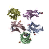
C: DNA (5'-D(P*DCP*DCP*DCP*DAP*DTP*DCP*DGP*DTP*DAP*DT)-3') D: DNA (5'-D(*DTP*DTP*DTP*DTP*DAP*DTP*DAP*DCP*DGP*DAP*DTP*DGP*DGP*DG)-3') A: DNA polymerase III subunit beta B: DNA polymerase III subunit beta hetero molecules
DNA (5'-D(P*DCP*DCP*DCP*DAP*DTP*DCP*DGP*DTP*DAP*DT)-3')
Mass: 2979.968 Da / Num. of mol.: 1 / Source method: obtained synthetically Details: DNA labeled at 5' end by dye CY5 (labeled as residue name 5CY). Only half of dye is visible in density
#2: DNA chain
DNA (5'-D(*DTP*DTP*DTP*DTP*DAP*DTP*DAP*DCP*DGP*DAP*DTP*DGP*DGP*DG)-3')
Mass: 4325.825 Da / Num. of mol.: 1 / Source method: obtained synthetically
#3: Protein
DNApolymeraseIIIsubunitbeta / DNA polymerase III holoenzyme / E.C.2.7.7.7
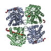
Mass: 40630.508 Da / Num. of mol.: 2 Source method: isolated from a genetically manipulated source Source: (gene. exp.) Escherichia coli (E. coli) / Gene: dnaN / Plasmid: pET / Species (production host): Escherichia coli / Production host: Escherichia coli BL21 (bacteria) / Strain (production host): BL21 / References: UniProt: P0A988, DNA-directed DNA polymerase
In the structure databanks used in Yorodumi, some data are registered as the other names, "COVID-19 virus" and "2019-nCoV". Here are the details of the virus and the list of structure data.
Jan 31, 2019. EMDB accession codes are about to change! (news from PDBe EMDB page)
EMDB accession codes are about to change! (news from PDBe EMDB page)
The allocation of 4 digits for EMDB accession codes will soon come to an end. Whilst these codes will remain in use, new EMDB accession codes will include an additional digit and will expand incrementally as the available range of codes is exhausted. The current 4-digit format prefixed with “EMD-” (i.e. EMD-XXXX) will advance to a 5-digit format (i.e. EMD-XXXXX), and so on. It is currently estimated that the 4-digit codes will be depleted around Spring 2019, at which point the 5-digit format will come into force.
The EM Navigator/Yorodumi systems omit the EMD- prefix.
Related info.:Q: What is EMD? / ID/Accession-code notation in Yorodumi/EM Navigator
Yorodumi is a browser for structure data from EMDB, PDB, SASBDB, etc.
This page is also the successor to EM Navigator detail page, and also detail information page/front-end page for Omokage search.
The word "yorodu" (or yorozu) is an old Japanese word meaning "ten thousand". "mi" (miru) is to see.
Related info.:EMDB / PDB / SASBDB / Comparison of 3 databanks / Yorodumi Search / Aug 31, 2016. New EM Navigator & Yorodumi / Yorodumi Papers / Jmol/JSmol / Function and homology information / Changes in new EM Navigator and Yorodumi
 Movie
Movie Controller


 Open data
 Basic information
 Components
 Keywords
 TRANSFERASE / TRANSCRIPTION/DNA / beta subunit /
  Function and homology information
 Authors
 Citation
 Structure visualization
 Downloads & links
 Other downloads
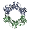
 PDBj
PDBj


 Assembly
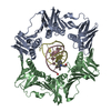
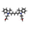
 Mass: 471.654 Da / Num. of mol.: 1 / Source method: obtained synthetically / Formula: C31H39N2O2
 Mass: 18.015 Da / Num. of mol.: 604 / Source method: isolated from a natural source / Formula: H2O
 Sample preparation
 / Beamline: X4A / Wavelength: 0.959 Å
 Processing