 Movie
Movie Controller

[English] 日本語
 Yorodumi
- PDB-3bef: Crystal structure of thrombin bound to the extracellular fragment... -
+ Open data

- Basic information
| Entry | Database: PDB / ID: 3bef | ||||||
|---|---|---|---|---|---|---|---|
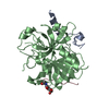
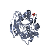
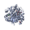
| Title | Crystal structure of thrombin bound to the extracellular fragment of PAR1 | ||||||
 Components Components |
| ||||||
 Keywords Keywords |  HYDROLASE / Serine protease / Acute phase / Blood coagulation / Cleavage on pair of basic residues / Disease mutation / Gamma-carboxyglutamic acid / Glycoprotein / Kringle / Secreted / Zymogen / G-protein coupled receptor / Membrane / Phosphoprotein / Receptor / Transducer / Transmembrane HYDROLASE / Serine protease / Acute phase / Blood coagulation / Cleavage on pair of basic residues / Disease mutation / Gamma-carboxyglutamic acid / Glycoprotein / Kringle / Secreted / Zymogen / G-protein coupled receptor / Membrane / Phosphoprotein / Receptor / Transducer / Transmembrane | ||||||
| Function / homology |  Function and homology information Function and homology informationnegative regulation of renin secretion into blood stream / dendritic cell homeostasis / negative regulation of glomerular filtration / thrombin-activated receptor activity / establishment of synaptic specificity at neuromuscular junction / platelet dense tubular network / cell-cell junction maintenance / regulation of interleukin-1 beta production / connective tissue replacement involved in inflammatory response wound healing / platelet dense granule organization ...negative regulation of renin secretion into blood stream / dendritic cell homeostasis / negative regulation of glomerular filtration / thrombin-activated receptor activity / establishment of synaptic specificity at neuromuscular junction / platelet dense tubular network / cell-cell junction maintenance / regulation of interleukin-1 beta production / connective tissue replacement involved in inflammatory response wound healing / platelet dense granule organization / trans-synaptic signaling by endocannabinoid, modulating synaptic transmission / regulation of sensory perception of pain / positive regulation of smooth muscle contraction / positive regulation of calcium ion transport / protein kinase C-activating G protein-coupled receptor signaling pathway / positive regulation of Rho protein signal transduction / positive regulation of lipid kinase activity / positive regulation of phospholipase C-activating G protein-coupled receptor signaling pathway / cytolysis by host of symbiont cells / thrombospondin receptor activity / Defective factor XII causes hereditary angioedema / thrombin / neutrophil-mediated killing of gram-negative bacterium / regulation of blood coagulation / ligand-gated ion channel signaling pathway / Defective F8 cleavage by thrombin / Platelet Aggregation (Plug Formation) / positive regulation of cysteine-type endopeptidase activity involved in apoptotic process / negative regulation of platelet activation / negative regulation of astrocyte differentiation / G-protein alpha-subunit binding / negative regulation of cytokine production involved in inflammatory response / anatomical structure morphogenesis / positive regulation of collagen biosynthetic process / positive regulation of blood coagulation / negative regulation of fibrinolysis / Gamma-carboxylation of protein precursors / Transport of gamma-carboxylated protein precursors from the endoplasmic reticulum to the Golgi apparatus / Common Pathway of Fibrin Clot Formation / Removal of aminoterminal propeptides from gamma-carboxylated proteins / homeostasis of number of cells within a tissue / release of sequestered calcium ion into cytosol / positive regulation of vasoconstriction / regulation of cytosolic calcium ion concentration / fibrinolysis / Intrinsic Pathway of Fibrin Clot Formation / Peptide ligand-binding receptors / positive regulation of release of sequestered calcium ion into cytosol / Regulation of Complement cascade / positive regulation of interleukin-8 production / caveola / G protein-coupled receptor activity / acute-phase response / Cell surface interactions at the vascular wall / lipopolysaccharide binding / negative regulation of proteolysis / positive regulation of receptor signaling pathway via JAK-STAT / growth factor activity / neuromuscular junction / positive regulation of insulin secretion / platelet activation / positive regulation of GTPase activity / response to wounding / positive regulation of interleukin-6 production / Golgi lumen / positive regulation of protein localization to nucleus / Regulation of Insulin-like Growth Factor (IGF) transport and uptake by Insulin-like Growth Factor Binding Proteins (IGFBPs) / G-protein beta-subunit binding / activation of cysteine-type endopeptidase activity involved in apoptotic process / positive regulation of reactive oxygen species metabolic process / blood coagulation / antimicrobial humoral immune response mediated by antimicrobial peptide / Thrombin signalling through proteinase activated receptors (PARs) / late endosome / heparin binding / phospholipase C-activating G protein-coupled receptor signaling pathway / positive regulation of cytosolic calcium ion concentration / regulation of cell shape / postsynaptic membrane / positive regulation of cell growth / G alpha (q) signalling events / collagen-containing extracellular matrix / blood microparticle / positive regulation of canonical NF-kappaB signal transduction / negative regulation of neuron apoptotic process / response to lipopolysaccharide / positive regulation of MAPK cascade / positive regulation of ERK1 and ERK2 cascade / cell surface receptor signaling pathway / positive regulation of phosphatidylinositol 3-kinase/protein kinase B signal transduction / early endosome / positive regulation of cell migration / inflammatory response / positive regulation of protein phosphorylation / G protein-coupled receptor signaling pathway / endoplasmic reticulum lumen / negative regulation of cell population proliferation / signaling receptor binding / serine-type endopeptidase activity / calcium ion bindingSimilarity search - Function | ||||||
| Biological species |  Homo sapiens (human) Homo sapiens (human) | ||||||
| Method | X-RAY DIFFRACTION / SYNCHROTRON / MOLECULAR REPLACEMENT / Resolution: 2.2 Å | ||||||
 Authors Authors | Gandhi, P.S. / Bah, A. / Chen, Z. / Mathews, F.S. / Di Cera, E. | ||||||
 Citation Citation | Journal: Proc.Natl.Acad.Sci.Usa / Year: 2008 Title: Structural identification of the pathway of long-range communication in an allosteric enzyme. Authors: Gandhi, P.S. / Chen, Z. / Mathews, F.S. / Di Cera, E. #1: Journal: J.Biol.Chem. / Year: 2004Title: Molecular dissection of Na+ binding to thrombin Authors: Pineda, A.O. / Carrell, C.J. / Bush, L.A. / Prasad, S. / Caccia, S. / Chen, Z. / Mathews, F.S. / Di Cera, E. #2: Journal: J.Biol.Chem. / Year: 2006Title: Crystal Structure of Thrombin in a Self-inhibited Conformation Authors: Pineda, A.O. / Chen, Z. / Mathews, F.S. / Di Cera, E. | ||||||
| History |
|
- Structure visualization
| Structure viewer | Molecule: MolmilJmol/JSmol |
|---|
- Downloads & links
-Download
| PDBx/mmCIF format | 3bef.cif.gz | 140.6 KB | Display | PDBx/mmCIF format |
|---|---|---|---|---|
| PDB format | pdb3bef.ent.gz | 109 KB | Display | PDB format |
| PDBx/mmJSON format | 3bef.json.gz | Tree view | PDBx/mmJSON format | |
| Others |  Other downloads Other downloads |
-Validation report
| Arichive directory | https://data.pdbj.org/pub/pdb/validation_reports/be/3befftp://data.pdbj.org/pub/pdb/validation_reports/be/3bef | HTTPS FTP |
|---|
-Related structure data
| Related structure data |  3beiC  1shhS S: Starting model for refinement C: citing same article ( |
|---|---|
| Similar structure data |


-Links
 PDBj
PDBj


- Assembly
| Deposited unit | 
| ||||||||
|---|---|---|---|---|---|---|---|---|---|
| 1 | 
| ||||||||
| 2 | 
| ||||||||
| Unit cell |
|
-Components
| #1: Protein/peptide | Thrombin / COAGULATION FACTOR II Mass: 5367.890 Da / Num. of mol.: 2 / Fragment: THROMBIN LIGHT CHAIN / Mutation: D102N Source method: isolated from a genetically manipulated source Source: (gene. exp.) Homo sapiens (human) / Gene: F2 / Production host:   Cricetulus griseus (Chinese hamster) / References: UniProt: P00734, thrombin Cricetulus griseus (Chinese hamster) / References: UniProt: P00734, thrombin#2: Protein | Thrombin / COAGULATION FACTOR IIMass: 29779.234 Da / Num. of mol.: 2 / Fragment: THROMBIN HEAVY CHAIN Source method: isolated from a genetically manipulated source Source: (gene. exp.) Homo sapiens (human) / Gene: F2 / Production host: Cricetulus griseus (Chinese hamster) / References: UniProt: P00734, thrombin#3: Protein/peptide | Mass: 1228.286 Da / Num. of mol.: 2 / Fragment: UNP residues 49-57 Source method: isolated from a genetically manipulated source Source: (gene. exp.) Homo sapiens (human) / Gene: F2R, CF2R, PAR1, TR / Production host: Cricetulus griseus (Chinese hamster) / References: UniProt: P25116#4: Sugar | N-Acetylglucosamine  Type: D-saccharide, beta linking / Mass: 221.208 Da / Num. of mol.: 2 Type: D-saccharide, beta linking / Mass: 221.208 Da / Num. of mol.: 2Source method: isolated from a genetically manipulated source Formula: C8H15NO6 #5: Water | ChemComp-HOH / | Water Mass: 18.015 Da / Num. of mol.: 229 / Source method: isolated from a natural source / Formula: H2O Mass: 18.015 Da / Num. of mol.: 229 / Source method: isolated from a natural source / Formula: H2O |
|---|
-Experimental details
-Experiment
| Experiment | Method: X-RAY DIFFRACTION / Number of used crystals: 1 |
|---|
- Sample preparation
| Crystal | Density Matthews: 2.54 Å3/Da / Density % sol: 51.54 % |
|---|---|
| Crystal grow | Temperature: 285 K / Method: vapor diffusion, hanging drop / pH: 6.5 Details: 100mM MES, 30% PEG 4000, pH 6.5, VAPOR DIFFUSION, HANGING DROP, temperature 285K |
-Data collection
| Diffraction | Mean temperature: 100 K |
|---|---|
| Diffraction source | Source: SYNCHROTRON / Site: APS  / Beamline: 14-BM-C / Wavelength: 0.9 Å / Beamline: 14-BM-C / Wavelength: 0.9 Å |
| Detector | Type: ADSC QUANTUM 315 / Detector: CCD / Date: Dec 3, 2006 |
| Radiation | Monochromator: APS 14_BM_C / Protocol: SINGLE WAVELENGTH / Monochromatic (M) / Laue (L): M / Scattering type: x-ray |
| Radiation wavelength | Wavelength: 0.9 Å / Relative weight: 1 |
| Reflection | Resolution: 2.2→40 Å / Num. all: 36229 / Num. obs: 34055 / % possible obs: 94 % / Observed criterion σ(F): -1 / Observed criterion σ(I): -1 / Redundancy: 2.4 % / Biso Wilson estimate: 14.9 Å2 / Rmerge(I) obs: 0.061 / Net I/σ(I): 12.3 |
| Reflection shell | Resolution: 2.2→2.28 Å / Redundancy: 2 % / Rmerge(I) obs: 0.221 / Mean I/σ(I) obs: 3.1 / Num. unique all: 3155 / % possible all: 87.7 |
- Processing
| Software |
| |||||||||||||||||||||||||
|---|---|---|---|---|---|---|---|---|---|---|---|---|---|---|---|---|---|---|---|---|---|---|---|---|---|---|
| Refinement | Method to determine structure: MOLECULAR REPLACEMENT Starting model: PDB ENTry 1SHH Resolution: 2.2→28 Å / Rfactor Rfree error: 0.006 / Data cutoff high absF: 284604.08 / Data cutoff low absF: 0 / Isotropic thermal model: RESTRAINED / Cross valid method: THROUGHOUT / σ(F): 0 / σ(I): 0 / Stereochemistry target values: Engh & Huber
| |||||||||||||||||||||||||
| Displacement parameters | Biso mean: 37.9 Å2 | |||||||||||||||||||||||||
| Refine analyze |
| |||||||||||||||||||||||||
| Refinement step | Cycle: LAST / Resolution: 2.2→28 Å
| |||||||||||||||||||||||||
| Refine LS restraints |
| |||||||||||||||||||||||||
| LS refinement shell | Resolution: 2.2→2.34 Å / Rfactor Rfree error: 0.018 / Total num. of bins used: 6
|
