 Movie
Movie Controller

[English] 日本語
 Yorodumi
- PDB-3a9k: Crystal structure of the mouse TAB3-NZF in complex with Lys63-lin... -
+ Open data

- Basic information
| Entry | Database: PDB / ID: 3a9k | ||||||
|---|---|---|---|---|---|---|---|
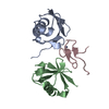
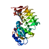
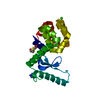
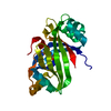
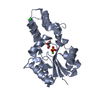
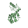
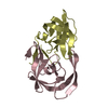
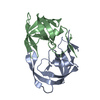
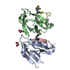
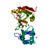
| Title | Crystal structure of the mouse TAB3-NZF in complex with Lys63-linked di-ubiquitin | ||||||
 Components Components |
| ||||||
 Keywords Keywords |  SIGNALING PROTEIN/METAL BINDING PROTEIN / protein complex / Cytoplasm / Isopeptide bond / Metal-binding / Zinc / Zinc-finger / SIGNALING PROTEIN-METAL BINDING PROTEIN complex SIGNALING PROTEIN/METAL BINDING PROTEIN / protein complex / Cytoplasm / Isopeptide bond / Metal-binding / Zinc / Zinc-finger / SIGNALING PROTEIN-METAL BINDING PROTEIN complex | ||||||
| Function / homology |  Function and homology information: / : / Formation of a pool of free 40S subunits / APC/C:Cdc20 mediated degradation of Cyclin B / SCF-beta-TrCP mediated degradation of Emi1 / APC-Cdc20 mediated degradation of Nek2A / ER Quality Control Compartment (ERQC) / Regulation of PTEN localization / Downregulation of ERBB2:ERBB3 signaling / SRP-dependent cotranslational protein targeting to membrane ...: / : / Formation of a pool of free 40S subunits / APC/C:Cdc20 mediated degradation of Cyclin B / SCF-beta-TrCP mediated degradation of Emi1 / APC-Cdc20 mediated degradation of Nek2A / ER Quality Control Compartment (ERQC) / Regulation of PTEN localization / Downregulation of ERBB2:ERBB3 signaling / SRP-dependent cotranslational protein targeting to membrane / SMAD2/SMAD3:SMAD4 heterotrimer regulates transcription / Major pathway of rRNA processing in the nucleolus and cytosol / IRAK2 mediated activation of TAK1 complex / Negative regulation of FLT3 / Downregulation of SMAD2/3:SMAD4 transcriptional activity / PTK6 Regulates RTKs and Their Effectors AKT1 and DOK1 / Regulation of expression of SLITs and ROBOs / Nonsense Mediated Decay (NMD) independent of the Exon Junction Complex (EJC) / Gap-filling DNA repair synthesis and ligation in GG-NER / Fanconi Anemia Pathway / Endosomal Sorting Complex Required For Transport (ESCRT) / Downregulation of TGF-beta receptor signaling / TGF-beta receptor signaling in EMT (epithelial to mesenchymal transition) / Synthesis of active ubiquitin: roles of E1 and E2 enzymes / IRAK1 recruits IKK complex / IRAK1 recruits IKK complex upon TLR7/8 or 9 stimulation / Downregulation of ERBB4 signaling / E3 ubiquitin ligases ubiquitinate target proteins / Alpha-protein kinase 1 signaling pathway / Stabilization of p53 / NOTCH3 Activation and Transmission of Signal to the Nucleus / Negative regulators of DDX58/IFIH1 signaling / Pexophagy / Regulation of NF-kappa B signaling / Nonsense Mediated Decay (NMD) enhanced by the Exon Junction Complex (EJC) / JNK (c-Jun kinases) phosphorylation and activation mediated by activated human TAK1 / Translesion synthesis by REV1 / Negative regulation of FGFR3 signaling / Negative regulation of FGFR4 signaling / Translesion synthesis by POLK / Negative regulation of FGFR1 signaling / Negative regulation of FGFR2 signaling / Regulation of TP53 Activity through Methylation / TRAF6-mediated induction of TAK1 complex within TLR4 complex / IRAK2 mediated activation of TAK1 complex upon TLR7/8 or 9 stimulation / Regulation of BACH1 activity / NRIF signals cell death from the nucleus / Translesion synthesis by POLI / Recognition of DNA damage by PCNA-containing replication complex / p75NTR recruits signalling complexes / HDR through Homologous Recombination (HRR) / Interferon alpha/beta signaling / Regulation of innate immune responses to cytosolic DNA / Negative regulation of MAPK pathway / Spry regulation of FGF signaling / Regulation of TP53 Degradation / Translesion Synthesis by POLH / Activated NOTCH1 Transmits Signal to the Nucleus / PINK1-PRKN Mediated Mitophagy / DNA Damage Recognition in GG-NER / Formation of TC-NER Pre-Incision Complex / Negative regulation of MET activity / Autodegradation of Cdh1 by Cdh1:APC/C / APC/C:Cdc20 mediated degradation of Securin / Termination of translesion DNA synthesis / Ubiquitin Mediated Degradation of Phosphorylated Cdc25A / Ubiquitin-dependent degradation of Cyclin D / Activation of IRF3, IRF7 mediated by TBK1, IKKε (IKBKE) / Inactivation of CSF3 (G-CSF) signaling / Senescence-Associated Secretory Phenotype (SASP) / AUF1 (hnRNP D0) binds and destabilizes mRNA / TNFR1-induced NF-kappa-B signaling pathway / Josephin domain DUBs / Dual Incision in GG-NER / Regulation of FZD by ubiquitination / Downregulation of ERBB2 signaling / Dual incision in TC-NER / IKK complex recruitment mediated by RIP1 / Cdc20:Phospho-APC/C mediated degradation of Cyclin A / SCF(Skp2)-mediated degradation of p27/p21 / Oncogene Induced Senescence / Assembly of the pre-replicative complex / CDK-mediated phosphorylation and removal of Cdc6 / TCF dependent signaling in response to WNT / N-glycan trimming in the ER and Calnexin/Calreticulin cycle / Formation of Incision Complex in GG-NER / Metalloprotease DUBs / Gap-filling DNA repair synthesis and ligation in TC-NER / L13a-mediated translational silencing of Ceruloplasmin expression / Degradation of AXIN / Regulation of TNFR1 signaling / GTP hydrolysis and joining of the 60S ribosomal subunit / EGFR downregulation / Autodegradation of the E3 ubiquitin ligase COP1 / Regulation of necroptotic cell death / MAP3K8 (TPL2)-dependent MAPK1/3 activation / G2/M Checkpoints / Asymmetric localization of PCP proteins / APC/C:Cdh1 mediated degradation of Cdc20 and other APC/C:Cdh1 targeted proteins in late mitosis/early G1 / Regulation of RUNX3 expression and activity Function and homology information: / : / Formation of a pool of free 40S subunits / APC/C:Cdc20 mediated degradation of Cyclin B / SCF-beta-TrCP mediated degradation of Emi1 / APC-Cdc20 mediated degradation of Nek2A / ER Quality Control Compartment (ERQC) / Regulation of PTEN localization / Downregulation of ERBB2:ERBB3 signaling / SRP-dependent cotranslational protein targeting to membrane ...: / : / Formation of a pool of free 40S subunits / APC/C:Cdc20 mediated degradation of Cyclin B / SCF-beta-TrCP mediated degradation of Emi1 / APC-Cdc20 mediated degradation of Nek2A / ER Quality Control Compartment (ERQC) / Regulation of PTEN localization / Downregulation of ERBB2:ERBB3 signaling / SRP-dependent cotranslational protein targeting to membrane / SMAD2/SMAD3:SMAD4 heterotrimer regulates transcription / Major pathway of rRNA processing in the nucleolus and cytosol / IRAK2 mediated activation of TAK1 complex / Negative regulation of FLT3 / Downregulation of SMAD2/3:SMAD4 transcriptional activity / PTK6 Regulates RTKs and Their Effectors AKT1 and DOK1 / Regulation of expression of SLITs and ROBOs / Nonsense Mediated Decay (NMD) independent of the Exon Junction Complex (EJC) / Gap-filling DNA repair synthesis and ligation in GG-NER / Fanconi Anemia Pathway / Endosomal Sorting Complex Required For Transport (ESCRT) / Downregulation of TGF-beta receptor signaling / TGF-beta receptor signaling in EMT (epithelial to mesenchymal transition) / Synthesis of active ubiquitin: roles of E1 and E2 enzymes / IRAK1 recruits IKK complex / IRAK1 recruits IKK complex upon TLR7/8 or 9 stimulation / Downregulation of ERBB4 signaling / E3 ubiquitin ligases ubiquitinate target proteins / Alpha-protein kinase 1 signaling pathway / Stabilization of p53 / NOTCH3 Activation and Transmission of Signal to the Nucleus / Negative regulators of DDX58/IFIH1 signaling / Pexophagy / Regulation of NF-kappa B signaling / Nonsense Mediated Decay (NMD) enhanced by the Exon Junction Complex (EJC) / JNK (c-Jun kinases) phosphorylation and activation mediated by activated human TAK1 / Translesion synthesis by REV1 / Negative regulation of FGFR3 signaling / Negative regulation of FGFR4 signaling / Translesion synthesis by POLK / Negative regulation of FGFR1 signaling / Negative regulation of FGFR2 signaling / Regulation of TP53 Activity through Methylation / TRAF6-mediated induction of TAK1 complex within TLR4 complex / IRAK2 mediated activation of TAK1 complex upon TLR7/8 or 9 stimulation / Regulation of BACH1 activity / NRIF signals cell death from the nucleus / Translesion synthesis by POLI / Recognition of DNA damage by PCNA-containing replication complex / p75NTR recruits signalling complexes / HDR through Homologous Recombination (HRR) / Interferon alpha/beta signaling / Regulation of innate immune responses to cytosolic DNA / Negative regulation of MAPK pathway / Spry regulation of FGF signaling / Regulation of TP53 Degradation / Translesion Synthesis by POLH / Activated NOTCH1 Transmits Signal to the Nucleus / PINK1-PRKN Mediated Mitophagy / DNA Damage Recognition in GG-NER / Formation of TC-NER Pre-Incision Complex / Negative regulation of MET activity / Autodegradation of Cdh1 by Cdh1:APC/C / APC/C:Cdc20 mediated degradation of Securin / Termination of translesion DNA synthesis / Ubiquitin Mediated Degradation of Phosphorylated Cdc25A / Ubiquitin-dependent degradation of Cyclin D / Activation of IRF3, IRF7 mediated by TBK1, IKKε (IKBKE) / Inactivation of CSF3 (G-CSF) signaling / Senescence-Associated Secretory Phenotype (SASP) / AUF1 (hnRNP D0) binds and destabilizes mRNA / TNFR1-induced NF-kappa-B signaling pathway / Josephin domain DUBs / Dual Incision in GG-NER / Regulation of FZD by ubiquitination / Downregulation of ERBB2 signaling / Dual incision in TC-NER / IKK complex recruitment mediated by RIP1 / Cdc20:Phospho-APC/C mediated degradation of Cyclin A / SCF(Skp2)-mediated degradation of p27/p21 / Oncogene Induced Senescence / Assembly of the pre-replicative complex / CDK-mediated phosphorylation and removal of Cdc6 / TCF dependent signaling in response to WNT / N-glycan trimming in the ER and Calnexin/Calreticulin cycle / Formation of Incision Complex in GG-NER / Metalloprotease DUBs / Gap-filling DNA repair synthesis and ligation in TC-NER / L13a-mediated translational silencing of Ceruloplasmin expression / Degradation of AXIN / Regulation of TNFR1 signaling / GTP hydrolysis and joining of the 60S ribosomal subunit / EGFR downregulation / Autodegradation of the E3 ubiquitin ligase COP1 / Regulation of necroptotic cell death / MAP3K8 (TPL2)-dependent MAPK1/3 activation / G2/M Checkpoints / Asymmetric localization of PCP proteins / APC/C:Cdh1 mediated degradation of Cdc20 and other APC/C:Cdh1 targeted proteins in late mitosis/early G1 / Regulation of RUNX3 expression and activitySimilarity search - Function | ||||||
| Biological species |  Mus musculus (house mouse) Mus musculus (house mouse) | ||||||
| Method | X-RAY DIFFRACTION / SYNCHROTRON / MOLECULAR REPLACEMENT / Resolution: 1.4 Å | ||||||
 Authors Authors | Sato, Y. / Yoshikawa, A. / Yamashita, M. / Yamagata, A. / Fukai, S. | ||||||
 Citation Citation | Journal: Embo J. / Year: 2009 Title: Structural basis for specific recognition of Lys 63-linked polyubiquitin chains by NZF domains of TAB2 and TAB3 Authors: Sato, Y. / Yoshikawa, A. / Yamashita, M. / Yamagata, A. / Fukai, S. | ||||||
| History |
|
- Structure visualization
| Structure viewer | Molecule: MolmilJmol/JSmol |
|---|
- Downloads & links
-Download
| PDBx/mmCIF format | 3a9k.cif.gz | 90.7 KB | Display | PDBx/mmCIF format |
|---|---|---|---|---|
| PDB format | pdb3a9k.ent.gz | 68.6 KB | Display | PDB format |
| PDBx/mmJSON format | 3a9k.json.gz | Tree view | PDBx/mmJSON format | |
| Others |  Other downloads Other downloads |
-Validation report
| Arichive directory | https://data.pdbj.org/pub/pdb/validation_reports/a9/3a9kftp://data.pdbj.org/pub/pdb/validation_reports/a9/3a9k | HTTPS FTP |
|---|
-Related structure data
| Related structure data |  3a9jSC S: Starting model for refinement C: citing same article ( |
|---|---|
| Similar structure data |
-Links
 PDBj
PDBj


- Assembly
| Deposited unit | 
| ||||||||
|---|---|---|---|---|---|---|---|---|---|
| 1 |
| ||||||||
| Unit cell |
| ||||||||
| Details | CHAINS A AND B ARE COVALENTLY BONDED TO FORM A SINGLE MOLECULE. THIS MOLECULE FORM A HETERO DIMERIC COMPLEX WITH CHAIN C. |
-Components
| #1: Protein | Mass: 8604.845 Da / Num. of mol.: 1 / Mutation: K63R Source method: isolated from a genetically manipulated source Source: (gene. exp.) Mus musculus (house mouse) / Gene: Ubiquitin / Plasmid: pET26b / Production host:  Escherichia coli (E. coli) / Strain (production host): Rosetta (DE3) / References: UniProt: P62991, UniProt: P0CG50*PLUS Escherichia coli (E. coli) / Strain (production host): Rosetta (DE3) / References: UniProt: P62991, UniProt: P0CG50*PLUS |
|---|---|
| #2: Protein | Mass: 8691.918 Da / Num. of mol.: 1 / Mutation: 77D Source method: isolated from a genetically manipulated source Source: (gene. exp.) Mus musculus (house mouse) / Gene: Ubiquitin / Plasmid: pET26b / Production host: Escherichia coli (E. coli) / Strain (production host): Rosetta (DE3) / References: UniProt: P62991, UniProt: P0CG50*PLUS |
| #3: Protein/peptide | Mass: 3841.340 Da / Num. of mol.: 1 / Fragment: RanBP2-type, residues 688-716 Source method: isolated from a genetically manipulated source Source: (gene. exp.) Mus musculus (house mouse) / Gene: TAB3 (AMINO ACIDS 688 - 716) / Plasmid: pCold GST / Production host: Escherichia coli (E. coli) / Strain (production host): Rosetta (DE3) / References: UniProt: Q571K4 |
| #4: Chemical | ChemComp-ZN /   Mass: 65.409 Da / Num. of mol.: 1 / Source method: obtained synthetically / Formula: Zn Mass: 65.409 Da / Num. of mol.: 1 / Source method: obtained synthetically / Formula: Zn |
| #5: Water | ChemComp-HOH / Water Mass: 18.015 Da / Num. of mol.: 144 / Source method: isolated from a natural source / Formula: H2O Mass: 18.015 Da / Num. of mol.: 144 / Source method: isolated from a natural source / Formula: H2O |
-Experimental details
-Experiment
| Experiment | Method: X-RAY DIFFRACTION / Number of used crystals: 1 |
|---|
- Sample preparation
| Crystal | Density Matthews: 1.8 Å3/Da / Density % sol: 31.8 % |
|---|---|
| Crystal grow | Temperature: 293 K / Method: vapor diffusion, sitting drop / pH: 6.5 Details: 100mM Bis-Tris-HCl (pH 6.5), 21% PEG 3350, VAPOR DIFFUSION, SITTING DROP, temperature 293K |
-Data collection
| Diffraction | Mean temperature: 100 K |
|---|---|
| Diffraction source | Source: SYNCHROTRON / Site: SPring-8  / Beamline: BL41XU / Wavelength: 1 Å / Beamline: BL41XU / Wavelength: 1 Å |
| Detector | Type: RAYONIX MX225HE / Detector: CCD / Date: May 29, 2009 / Details: mirrors |
| Radiation | Protocol: SINGLE WAVELENGTH / Monochromatic (M) / Laue (L): M / Scattering type: x-ray |
| Radiation wavelength | Wavelength: 1 Å / Relative weight: 1 |
| Reflection | Resolution: 1.4→50 Å / Num. all: 30844 / Num. obs: 30844 / % possible obs: 97.8 % / Observed criterion σ(I): 0 / Rmerge(I) obs: 0.05 / Net I/σ(I): 36.8 |
| Reflection shell | Resolution: 1.4→1.42 Å / Rmerge(I) obs: 0.347 / Mean I/σ(I) obs: 2.78 / % possible all: 93.5 |
- Processing
| Software |
| ||||||||||||||||||||||||||||||||||||||||||||||||||||||||||||||||||||||||||||||||||||||||||||||||||||||||||||||||||||||||||||||||||||||||||||||||||||||||||||||||||||||||||
|---|---|---|---|---|---|---|---|---|---|---|---|---|---|---|---|---|---|---|---|---|---|---|---|---|---|---|---|---|---|---|---|---|---|---|---|---|---|---|---|---|---|---|---|---|---|---|---|---|---|---|---|---|---|---|---|---|---|---|---|---|---|---|---|---|---|---|---|---|---|---|---|---|---|---|---|---|---|---|---|---|---|---|---|---|---|---|---|---|---|---|---|---|---|---|---|---|---|---|---|---|---|---|---|---|---|---|---|---|---|---|---|---|---|---|---|---|---|---|---|---|---|---|---|---|---|---|---|---|---|---|---|---|---|---|---|---|---|---|---|---|---|---|---|---|---|---|---|---|---|---|---|---|---|---|---|---|---|---|---|---|---|---|---|---|---|---|---|---|---|---|---|
| Refinement | Method to determine structure: MOLECULAR REPLACEMENT Starting model: PDB ENTRY 3A9J Resolution: 1.4→25.72 Å / Cor.coef. Fo:Fc: 0.959 / Cor.coef. Fo:Fc free: 0.951 / SU B: 2.701 / SU ML: 0.048 / Cross valid method: THROUGHOUT / ESU R: 0.091 / ESU R Free: 0.074 / Stereochemistry target values: MAXIMUM LIKELIHOOD / Details: HYDROGENS HAVE BEEN ADDED IN THE RIDING POSITIONS
| ||||||||||||||||||||||||||||||||||||||||||||||||||||||||||||||||||||||||||||||||||||||||||||||||||||||||||||||||||||||||||||||||||||||||||||||||||||||||||||||||||||||||||
| Solvent computation | Ion probe radii: 0.8 Å / Shrinkage radii: 0.8 Å / VDW probe radii: 1.2 Å / Solvent model: MASK | ||||||||||||||||||||||||||||||||||||||||||||||||||||||||||||||||||||||||||||||||||||||||||||||||||||||||||||||||||||||||||||||||||||||||||||||||||||||||||||||||||||||||||
| Displacement parameters | Biso mean: 18.054 Å2
| ||||||||||||||||||||||||||||||||||||||||||||||||||||||||||||||||||||||||||||||||||||||||||||||||||||||||||||||||||||||||||||||||||||||||||||||||||||||||||||||||||||||||||
| Refinement step | Cycle: LAST / Resolution: 1.4→25.72 Å
| ||||||||||||||||||||||||||||||||||||||||||||||||||||||||||||||||||||||||||||||||||||||||||||||||||||||||||||||||||||||||||||||||||||||||||||||||||||||||||||||||||||||||||
| Refine LS restraints |
| ||||||||||||||||||||||||||||||||||||||||||||||||||||||||||||||||||||||||||||||||||||||||||||||||||||||||||||||||||||||||||||||||||||||||||||||||||||||||||||||||||||||||||
| LS refinement shell | Resolution: 1.401→1.437 Å / Total num. of bins used: 20
|
