 Movie
Movie Controller

[English] 日本語
 Yorodumi
- PDB-2zxr: Crystal structure of RecJ in complex with Mg2+ from Thermus therm... -
+ Open data

- Basic information
| Entry | Database: PDB / ID: 2zxr | ||||||
|---|---|---|---|---|---|---|---|
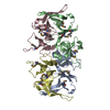
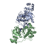
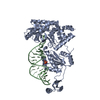
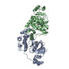
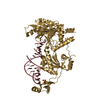
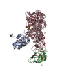
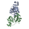
| Title | Crystal structure of RecJ in complex with Mg2+ from Thermus thermophilus HB8 | ||||||
 Components Components | Single-stranded DNA specific exonuclease RecJ | ||||||
 Keywords Keywords |  HYDROLASE / nuclease / single-stranded DNA / DNA repair / Exonuclease HYDROLASE / nuclease / single-stranded DNA / DNA repair / Exonuclease | ||||||
| Function / homology |  Function and homology informationexonuclease activity / Hydrolases; Acting on ester bonds / DNA binding / metal ion binding Function and homology informationexonuclease activity / Hydrolases; Acting on ester bonds / DNA binding / metal ion bindingSimilarity search - Function | ||||||
| Biological species |   Thermus thermophilus (bacteria) Thermus thermophilus (bacteria) | ||||||
| Method | X-RAY DIFFRACTION / SYNCHROTRON / MOLECULAR REPLACEMENT / Resolution: 2.15 Å | ||||||
 Authors Authors | Wakamatsu, T. / Kitamura, Y. / Nakagawa, N. / Masui, R. / Kuramitsu, S. | ||||||
 Citation Citation | Journal: J.Biol.Chem. / Year: 2010 Title: Structure of RecJ exonuclease defines its specificity for single-stranded DNA Authors: Wakamatsu, T. / Kitamura, Y. / Kotera, Y. / Nakagawa, N. / Kuramitsu, S. / Masui, R. | ||||||
| History |
|
- Structure visualization
| Structure viewer | Molecule: MolmilJmol/JSmol |
|---|
- Downloads & links
-Download
| PDBx/mmCIF format | 2zxr.cif.gz | 137.6 KB | Display | PDBx/mmCIF format |
|---|---|---|---|---|
| PDB format | pdb2zxr.ent.gz | 106.3 KB | Display | PDB format |
| PDBx/mmJSON format | 2zxr.json.gz | Tree view | PDBx/mmJSON format | |
| Others |  Other downloads Other downloads |
-Validation report
| Arichive directory | https://data.pdbj.org/pub/pdb/validation_reports/zx/2zxrftp://data.pdbj.org/pub/pdb/validation_reports/zx/2zxr | HTTPS FTP |
|---|
-Related structure data
| Related structure data |  2zxoSC 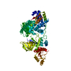 2zxpC S: Starting model for refinement C: citing same article ( |
|---|---|
| Similar structure data |


-Links
 PDBj
PDBj- Assembly
| Deposited unit | 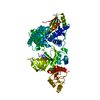
| ||||||||
|---|---|---|---|---|---|---|---|---|---|
| 1 |
| ||||||||
| Unit cell |
|
-Components
| #1: Protein | Mass: 72960.344 Da / Num. of mol.: 1 Source method: isolated from a genetically manipulated source Source: (gene. exp.) Thermus thermophilus (bacteria) / Strain: HB8 / Gene: recj, TTHA1167 / Plasmid: pET11a / Production host: Escherichia coli (E. coli) / Strain (production host): Rosetta2(DE3)pLysSReferences: UniProt: Q5SJ47, Hydrolases; Acting on ester bonds; Exodeoxyribonucleases producing 5'-phosphomonoesters |
|---|---|
| #2: Chemical | ChemComp-MG /   Mass: 24.305 Da / Num. of mol.: 1 / Source method: obtained synthetically / Formula: Mg Mass: 24.305 Da / Num. of mol.: 1 / Source method: obtained synthetically / Formula: Mg |
| #3: Water | ChemComp-HOH / Water Mass: 18.015 Da / Num. of mol.: 290 / Source method: isolated from a natural source / Formula: H2O Mass: 18.015 Da / Num. of mol.: 290 / Source method: isolated from a natural source / Formula: H2O |
-Experimental details
-Experiment
| Experiment | Method: X-RAY DIFFRACTION / Number of used crystals: 1 |
|---|
- Sample preparation
| Crystal | Density Matthews: 2.99 Å3/Da / Density % sol: 58.84 % |
|---|---|
| Crystal grow | Temperature: 293 K / Method: vapor diffusion, hanging drop / pH: 6.5 Details: 0.05M ADA, 6% PEG4000, 1% 2-propanol. The crystal was soaked in cryoprotecting solution containg 20% glycerol and 10mM magnesium sulfate, pH6.5, VAPOR DIFFUSION, HANGING DROP, temperature 293K |
-Data collection
| Diffraction | Mean temperature: 93 K |
|---|---|
| Diffraction source | Source: SYNCHROTRON / Site: SPring-8  / Beamline: BL26B2 / Wavelength: 1 Å / Beamline: BL26B2 / Wavelength: 1 Å |
| Detector | Type: RIGAKU RAXIS V / Detector: IMAGE PLATE / Date: Jul 18, 2008 |
| Radiation | Monochromator: Si double crystal / Protocol: SINGLE WAVELENGTH / Monochromatic (M) / Laue (L): M / Scattering type: x-ray |
| Radiation wavelength | Wavelength: 1 Å / Relative weight: 1 |
| Reflection | Resolution: 2.15→50 Å / Num. all: 48684 / Num. obs: 48684 / % possible obs: 98.5 % / Observed criterion σ(I): -3 / Redundancy: 17.2 % / Biso Wilson estimate: 20.1 Å2 / Rmerge(I) obs: 0.064 / Net I/σ(I): 47.2 |
| Reflection shell | Resolution: 2.15→2.23 Å / Redundancy: 3.9 % / Rmerge(I) obs: 0.276 / Mean I/σ(I) obs: 3.3 / Num. unique all: 837996 / % possible all: 86 |
- Processing
| Software |
| |||||||||||||||||||||||||
|---|---|---|---|---|---|---|---|---|---|---|---|---|---|---|---|---|---|---|---|---|---|---|---|---|---|---|
| Refinement | Method to determine structure: MOLECULAR REPLACEMENT Starting model: PDB ENTRY 2ZXO Resolution: 2.15→43.01 Å / Isotropic thermal model: restrained / Cross valid method: THROUGHOUT / σ(F): 0 / Stereochemistry target values: Engh & Huber
| |||||||||||||||||||||||||
| Displacement parameters | Biso mean: 34.9 Å2
| |||||||||||||||||||||||||
| Refine analyze |
| |||||||||||||||||||||||||
| Refinement step | Cycle: LAST / Resolution: 2.15→43.01 Å
| |||||||||||||||||||||||||
| Refine LS restraints |
| |||||||||||||||||||||||||
| LS refinement shell | Resolution: 2.15→2.28 Å / Rfactor Rfree error: 0.012
|
