 Movie
Movie Controller

[English] 日本語
 Yorodumi
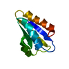
- PDB-2zqe: Crystal structure of the Smr domain of Thermus thermophilus MutS2 -
+ Open data

- Basic information
| Entry | Database: PDB / ID: 2zqe | ||||||
|---|---|---|---|---|---|---|---|
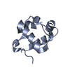
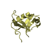
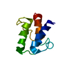
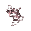
| Title | Crystal structure of the Smr domain of Thermus thermophilus MutS2 | ||||||
 Components Components | MutS2 protein | ||||||
 Keywords Keywords |  DNA BINDING PROTEIN / alpha/beta / ATP-binding / DNA-binding / Nucleotide-binding DNA BINDING PROTEIN / alpha/beta / ATP-binding / DNA-binding / Nucleotide-binding | ||||||
| Function / homology |  Function and homology information Function and homology informationmismatched DNA binding / negative regulation of DNA recombination / mismatch repair / endonuclease activity / Hydrolases; Acting on ester bonds / ATP hydrolysis activity / ATP bindingSimilarity search - Function | ||||||
| Biological species |   Thermus thermophilus (bacteria) Thermus thermophilus (bacteria) | ||||||
| Method | X-RAY DIFFRACTION / SYNCHROTRON / MOLECULAR REPLACEMENT / Resolution: 1.7 Å | ||||||
 Authors Authors | Fukui, K. / Kitamura, Y. / Nakagawa, N. / Masui, R. / Kuramitsu, S. | ||||||
 Citation Citation | Journal: J.Biol.Chem. / Year: 2008 Title: Crystal structure of MutS2 endonuclease domain and the mechanism of homologous recombination suppression Authors: Fukui, K. / Nakagawa, N. / Kitamura, Y. / Nishida, Y. / Masui, R. / Kuramitsu, S. | ||||||
| History |
|
- Structure visualization
| Structure viewer | Molecule: MolmilJmol/JSmol |
|---|
- Downloads & links
-Download
| PDBx/mmCIF format | 2zqe.cif.gz | 28.5 KB | Display | PDBx/mmCIF format |
|---|---|---|---|---|
| PDB format | pdb2zqe.ent.gz | 18.8 KB | Display | PDB format |
| PDBx/mmJSON format | 2zqe.json.gz | Tree view | PDBx/mmJSON format | |
| Others |  Other downloads Other downloads |
-Validation report
| Arichive directory | https://data.pdbj.org/pub/pdb/validation_reports/zq/2zqeftp://data.pdbj.org/pub/pdb/validation_reports/zq/2zqe | HTTPS FTP |
|---|
-Related structure data
| Related structure data |  2d9iS S: Starting model for refinement |
|---|---|
| Similar structure data |


-Links
 PDBj
PDBj- Assembly
| Deposited unit | 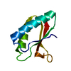
| ||||||||
|---|---|---|---|---|---|---|---|---|---|
| 1 |
| ||||||||
| Unit cell |
|
-Components
| #1: Protein | Mass: 8953.281 Da / Num. of mol.: 1 / Fragment: UNP Residue 663-744 Source method: isolated from a genetically manipulated source Source: (gene. exp.) Thermus thermophilus (bacteria) / Strain: HB8 / Gene: TTHA1645 / Plasmid: pET-11a / Production host: Escherichia coli (E. coli) / Strain (production host): BL21(DE3) / References: UniProt: Q5SHT5 |
|---|---|
| #2: Water | ChemComp-HOH / Water Mass: 18.015 Da / Num. of mol.: 104 / Source method: isolated from a natural source / Formula: H2O Mass: 18.015 Da / Num. of mol.: 104 / Source method: isolated from a natural source / Formula: H2O |
-Experimental details
-Experiment
| Experiment | Method: X-RAY DIFFRACTION / Number of used crystals: 1 |
|---|
- Sample preparation
| Crystal | Density Matthews: 2.23 Å3/Da / Density % sol: 44.96 % |
|---|---|
| Crystal grow | Temperature: 293 K / Method: vapor diffusion, hanging drop / pH: 3.5 Details: 0.1M Citric acid, 25% PEG 3350, pH 3.5, VAPOR DIFFUSION, HANGING DROP, temperature 293K |
-Data collection
| Diffraction | Mean temperature: 100 K |
|---|---|
| Diffraction source | Source: SYNCHROTRON / Site: SPring-8  / Beamline: BL26B2 / Wavelength: 1 Å / Beamline: BL26B2 / Wavelength: 1 Å |
| Detector | Type: RIGAKU JUPITER 210 / Detector: CCD / Date: Apr 11, 2008 |
| Radiation | Monochromator: transparent diamond double-crystal / Protocol: SINGLE WAVELENGTH / Monochromatic (M) / Laue (L): M / Scattering type: x-ray |
| Radiation wavelength | Wavelength: 1 Å / Relative weight: 1 |
| Reflection | Resolution: 1.7→50 Å / Num. obs: 9284 / % possible obs: 99.8 % / Redundancy: 6.3 % / Biso Wilson estimate: 11.8 Å2 / Rmerge(I) obs: 0.047 / Net I/σ(I): 36.4 |
| Reflection shell | Resolution: 1.7→1.76 Å / Redundancy: 6.3 % / Rmerge(I) obs: 0.137 / Mean I/σ(I) obs: 13.1 / Num. unique all: 58499 / % possible all: 99.8 |
- Processing
| Software |
| ||||||||||||||||||||||||||||||||||||||||||||||||||||||||||||||||||||||||||||||||||||||||||
|---|---|---|---|---|---|---|---|---|---|---|---|---|---|---|---|---|---|---|---|---|---|---|---|---|---|---|---|---|---|---|---|---|---|---|---|---|---|---|---|---|---|---|---|---|---|---|---|---|---|---|---|---|---|---|---|---|---|---|---|---|---|---|---|---|---|---|---|---|---|---|---|---|---|---|---|---|---|---|---|---|---|---|---|---|---|---|---|---|---|---|---|
| Refinement | Method to determine structure: MOLECULAR REPLACEMENT Starting model: PDB ENTRY 2d9i Resolution: 1.7→7.9 Å / Cor.coef. Fo:Fc: 0.939 / Cor.coef. Fo:Fc free: 0.934 / SU B: 1.899 / SU ML: 0.066 / Cross valid method: THROUGHOUT / ESU R: 0.114 / ESU R Free: 0.112 / Stereochemistry target values: MAXIMUM LIKELIHOOD / Details: HYDROGENS HAVE BEEN ADDED IN THE RIDING POSITIONS
| ||||||||||||||||||||||||||||||||||||||||||||||||||||||||||||||||||||||||||||||||||||||||||
| Solvent computation | Ion probe radii: 0.8 Å / Shrinkage radii: 0.8 Å / VDW probe radii: 1.2 Å / Solvent model: MASK | ||||||||||||||||||||||||||||||||||||||||||||||||||||||||||||||||||||||||||||||||||||||||||
| Displacement parameters | Biso mean: 11.168 Å2
| ||||||||||||||||||||||||||||||||||||||||||||||||||||||||||||||||||||||||||||||||||||||||||
| Refinement step | Cycle: LAST / Resolution: 1.7→7.9 Å
| ||||||||||||||||||||||||||||||||||||||||||||||||||||||||||||||||||||||||||||||||||||||||||
| Refine LS restraints |
| ||||||||||||||||||||||||||||||||||||||||||||||||||||||||||||||||||||||||||||||||||||||||||
| LS refinement shell | Resolution: 1.702→1.744 Å / Total num. of bins used: 20
|
