 Movie
Movie Controller

[English] 日本語
 Yorodumi
- PDB-2yyt: Crystal structure of uncharacterized conserved protein from Geoba... -
+ Open data

- Basic information
| Entry | Database: PDB / ID: 2yyt | ||||||
|---|---|---|---|---|---|---|---|
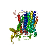
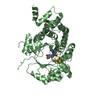
| Title | Crystal structure of uncharacterized conserved protein from Geobacillus kaustophilus | ||||||
 Components Components | Orotidine 5'-phosphate decarboxylase | ||||||
 Keywords Keywords | LYASE / TIM barrel / Structural Genomics / NPPSFA / National Project on Protein Structural and Functional Analyses / RIKEN Structural Genomics/Proteomics Initiative / RSGI | ||||||
| Function / homology |  Function and homology informationorotidine-5'-phosphate decarboxylase / orotidine-5'-phosphate decarboxylase activity / 'de novo' UMP biosynthetic process / 'de novo' pyrimidine nucleobase biosynthetic process Function and homology informationorotidine-5'-phosphate decarboxylase / orotidine-5'-phosphate decarboxylase activity / 'de novo' UMP biosynthetic process / 'de novo' pyrimidine nucleobase biosynthetic processSimilarity search - Function | ||||||
| Biological species |  Geobacillus kaustophilus (bacteria) Geobacillus kaustophilus (bacteria) | ||||||
| Method | X-RAY DIFFRACTION / SYNCHROTRON / MOLECULAR REPLACEMENT / Resolution: 2.3 Å | ||||||
 Authors Authors | Kanagawa, M. / Baba, S. / Nakamura, Y. / Bessho, Y. / Kuramitsu, S. / Yokoyama, S. / Kawai, G. / Sampei, G. / RIKEN Structural Genomics/Proteomics Initiative (RSGI) | ||||||
 Citation Citation | Journal: To be Published Title: Crystal structure of uncharacterized conserved protein from Geobacillus kaustophilus Authors: Kanagawa, M. / Baba, S. / Nakamura, Y. / Bessho, Y. / Kuramitsu, S. / Yokoyama, S. / Kawai, G. / Sampei, G. | ||||||
| History |
|
- Structure visualization
| Structure viewer | Molecule: MolmilJmol/JSmol |
|---|
- Downloads & links
-Download
| PDBx/mmCIF format | 2yyt.cif.gz | 179 KB | Display | PDBx/mmCIF format |
|---|---|---|---|---|
| PDB format | pdb2yyt.ent.gz | 143.3 KB | Display | PDB format |
| PDBx/mmJSON format | 2yyt.json.gz | Tree view | PDBx/mmJSON format | |
| Others |  Other downloads Other downloads |
-Validation report
| Arichive directory | https://data.pdbj.org/pub/pdb/validation_reports/yy/2yytftp://data.pdbj.org/pub/pdb/validation_reports/yy/2yyt | HTTPS FTP |
|---|
-Related structure data


-Links
 PDBj
PDBj
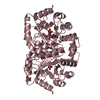
- Assembly
| Deposited unit | 
| ||||||||
|---|---|---|---|---|---|---|---|---|---|
| 1 | 
| ||||||||
| 2 | 
| ||||||||
| Unit cell |
|
-Components
| #1: Protein | / OMP decarboxylase / OMPDCase / OMPdecase Mass: 26982.936 Da / Num. of mol.: 4 Source method: isolated from a genetically manipulated source Source: (gene. exp.) Geobacillus kaustophilus (bacteria) / Strain: HTA426 / Plasmid: pET-HisTEV / Production host: Escherichia coli (E. coli) / Strain (production host): BL21-CodonPlus(DE3)-RIL-XReferences: UniProt: Q5L0U0, orotidine-5'-phosphate decarboxylase#2: Water | ChemComp-HOH / | Water Mass: 18.015 Da / Num. of mol.: 382 / Source method: isolated from a natural source / Formula: H2O Mass: 18.015 Da / Num. of mol.: 382 / Source method: isolated from a natural source / Formula: H2O |
|---|
-Experimental details
-Experiment
| Experiment | Method: X-RAY DIFFRACTION / Number of used crystals: 1 |
|---|
- Sample preparation
| Crystal | Density Matthews: 2.09 Å3/Da / Density % sol: 41.17 % Description: THE STRUCTURE FACTOR FILE CONTAINS FRIEDEL PAIRS. |
|---|---|
| Crystal grow | Temperature: 293 K / Method: vapor diffusion, sitting drop Details: 0.2M Sodium Sulfate decahydrate, 20% PEG 3350, VAPOR DIFFUSION, SITTING DROP, temperature 293K |
-Data collection
| Diffraction | Mean temperature: 100 K |
|---|---|
| Diffraction source | Source: SYNCHROTRON / Site: SPring-8  / Beamline: BL26B2 / Wavelength: 1 Å / Beamline: BL26B2 / Wavelength: 1 Å |
| Detector | Type: RIGAKU JUPITER 210 / Detector: CCD / Date: Oct 7, 2006 |
| Radiation | Monochromator: SI Double-Crystal / Protocol: SINGLE WAVELENGTH / Monochromatic (M) / Laue (L): M / Scattering type: x-ray |
| Radiation wavelength | Wavelength: 1 Å / Relative weight: 1 |
| Reflection | Resolution: 1.8→50 Å / Num. obs: 141451 / % possible obs: 100 % / Redundancy: 2.7 % / Biso Wilson estimate: 14.8 Å2 / Rsym value: 0.062 |
- Processing
| Software |
| ||||||||||||||||||||
|---|---|---|---|---|---|---|---|---|---|---|---|---|---|---|---|---|---|---|---|---|---|
| Refinement | Method to determine structure: MOLECULAR REPLACEMENT / Resolution: 2.3→50 Å / Rfactor Rfree error: 0.004 / Data cutoff high absF: 451132.4 / Data cutoff low absF: 0 / Isotropic thermal model: RESTRAINED / Cross valid method: THROUGHOUT / σ(F): 0 / Details: THE STRUCTURE FACTOR FILE CONTAINS FRIEDEL PAIRS.
| ||||||||||||||||||||
| Solvent computation | Solvent model: FLAT MODEL / Bsol: 44.0211 Å2 / ksol: 0.36303 e/Å3 | ||||||||||||||||||||
| Displacement parameters | Biso mean: 20.4 Å2
| ||||||||||||||||||||
| Refine analyze |
| ||||||||||||||||||||
| Refinement step | Cycle: LAST / Resolution: 2.3→50 Å
| ||||||||||||||||||||
| Refine LS restraints |
| ||||||||||||||||||||
| LS refinement shell | Resolution: 2.3→2.44 Å / Rfactor Rfree error: 0.01 / Total num. of bins used: 6
| ||||||||||||||||||||
| Xplor file |
|
