- PDB-2yye: Crystal structure of selenophosphate synthetase from Aquifex aeol... -
+
Open data
ID or keywords:
Loading...
-
Basic information
Entry
Database: PDB / ID: 2yye
Title
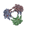
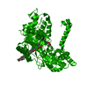
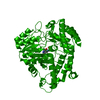
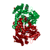
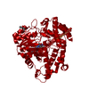
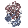
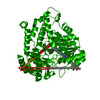
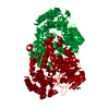
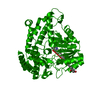
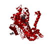
Crystal structure of selenophosphate synthetase from Aquifex aeolicus complexed with AMPCPP
Components
Selenide, water dikinase
Keywords
TRANSFERASE / Full-length selenophosphate synthetase complex with ATP-analog / Structural Genomics / NPPSFA / National Project on Protein Structural and Functional Analyses / RIKEN Structural Genomics/Proteomics Initiative / RSGI
Function / homology
Function and homology information
selenide, water dikinase / selenide, water dikinase activity / selenocysteine biosynthetic process / phosphorylation / magnesium ion binding / ATP binding / cytoplasm Similarity search - Function
Selenophosphate synthetase, class I / Selenophosphate synthetase / Phosphoribosyl-aminoimidazole Synthetase; Chain A, domain 2 / PurM-like C-terminal domain / PurM-like, N-terminal domain / PurM-like, N-terminal domain / AIR synthase related protein, N-terminal domain / PurM-like, C-terminal domain / PurM-like, C-terminal domain superfamily / PurM-like, N-terminal domain superfamily ...Selenophosphate synthetase, class I / Selenophosphate synthetase / Phosphoribosyl-aminoimidazole Synthetase; Chain A, domain 2 / PurM-like C-terminal domain / PurM-like, N-terminal domain / PurM-like, N-terminal domain / AIR synthase related protein, N-terminal domain / PurM-like, C-terminal domain / PurM-like, C-terminal domain superfamily / PurM-like, N-terminal domain superfamily / AIR synthase related protein, C-terminal domain / 60s Ribosomal Protein L30; Chain: A; / Alpha-Beta Complex / 2-Layer Sandwich / Alpha Beta Similarity search - Domain/homology
DIPHOSPHOMETHYLPHOSPHONIC ACID ADENOSYL ESTER / : / PHOSPHATE ION / Selenide, water dikinase Similarity search - Component
Resolution: 2.1→42.47 Å / Rfactor Rfree error: 0.005 / Data cutoff high absF: 2374350.3 / Data cutoff low absF: 0 / Isotropic thermal model: RESTRAINED / Cross valid method: THROUGHOUT / σ(F): 0 Details: We replaced selenocysteine 13 by Cys to facilitate the protein expression
In the structure databanks used in Yorodumi, some data are registered as the other names, "COVID-19 virus" and "2019-nCoV". Here are the details of the virus and the list of structure data.
Jan 31, 2019. EMDB accession codes are about to change! (news from PDBe EMDB page)
EMDB accession codes are about to change! (news from PDBe EMDB page)
The allocation of 4 digits for EMDB accession codes will soon come to an end. Whilst these codes will remain in use, new EMDB accession codes will include an additional digit and will expand incrementally as the available range of codes is exhausted. The current 4-digit format prefixed with “EMD-” (i.e. EMD-XXXX) will advance to a 5-digit format (i.e. EMD-XXXXX), and so on. It is currently estimated that the 4-digit codes will be depleted around Spring 2019, at which point the 5-digit format will come into force.
The EM Navigator/Yorodumi systems omit the EMD- prefix.
Related info.:Q: What is EMD? / ID/Accession-code notation in Yorodumi/EM Navigator
Yorodumi is a browser for structure data from EMDB, PDB, SASBDB, etc.
This page is also the successor to EM Navigator detail page, and also detail information page/front-end page for Omokage search.
The word "yorodu" (or yorozu) is an old Japanese word meaning "ten thousand". "mi" (miru) is to see.
Related info.:EMDB / PDB / SASBDB / Comparison of 3 databanks / Yorodumi Search / Aug 31, 2016. New EM Navigator & Yorodumi / Yorodumi Papers / Jmol/JSmol / Function and homology information / Changes in new EM Navigator and Yorodumi
 Movie
Movie Controller

 Yorodumi
 Open data
 Basic information
 Components
 Keywords
 Function and homology information

 Authors
 Citation
 Structure visualization
 Downloads & links
 Other downloads
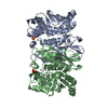


 PDBj
PDBj


 Assembly
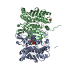

 Mass: 58.933 Da / Num. of mol.: 10 / Source method: obtained synthetically / Formula: Co
 Mass: 505.208 Da / Num. of mol.: 2 / Source method: obtained synthetically / Formula: C11H18N5O12P3 / Comment: AMP-CPP, energy-carrying molecule analogue*YM
 Mass: 94.971 Da / Num. of mol.: 1 / Source method: obtained synthetically / Formula: PO4
 Mass: 18.015 Da / Num. of mol.: 279 / Source method: isolated from a natural source / Formula: H2O
 Sample preparation
 / Beamline: BL41XU / Wavelength: 1 Å
 Processing