- PDB-2yiu: X-ray structure of the dimeric cytochrome BC1 complex from the so... -
+
Open data
ID or keywords:
Loading...
-
Basic information
Entry
Database: PDB / ID: 2yiu
Title
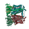
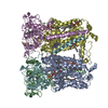
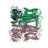
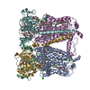
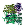
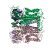
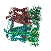
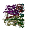
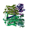
X-ray structure of the dimeric cytochrome BC1 complex from the soil bacterium paracoccus denitrificans at 2.7 angstrom resolution
Components
CYTOCHROME B
CYTOCHROME C1, HEME PROTEIN
UBIQUINOL-CYTOCHROME C REDUCTASE IRON-SULFUR SUBUNIT
Keywords
OXIDOREDUCTASE
Function / homology
Function and homology information
respiratory chain complex III / quinol-cytochrome-c reductase / ubiquinol-cytochrome-c reductase activity / respirasome / respiratory electron transport chain / 2 iron, 2 sulfur cluster binding / electron transfer activity / membrane => GO:0016020 / heme binding / metal ion binding / plasma membrane Similarity search - Function
Ubiquitinol-cytochrome C reductase, Fe-S subunit, TAT signal / Ubiquitinol-cytochrome C reductase Fe-S subunit TAT signal / Single helix bin / Cytochrome Bc1 Complex; Chain C / Cytochrome Bc1 Complex; Chain C / Cytochrome c1, transmembrane anchor, C-terminal / Rieske Iron-sulfur Protein / Rieske [2Fe-2S] iron-sulphur domain / 3-layer Sandwich / Cytochrome b ...Ubiquitinol-cytochrome C reductase, Fe-S subunit, TAT signal / Ubiquitinol-cytochrome C reductase Fe-S subunit TAT signal / Single helix bin / Cytochrome Bc1 Complex; Chain C / Cytochrome Bc1 Complex; Chain C / Cytochrome c1, transmembrane anchor, C-terminal / Rieske Iron-sulfur Protein / Rieske [2Fe-2S] iron-sulphur domain / 3-layer Sandwich / Cytochrome b / Ubiquinol-cytochrome c reductase, iron-sulphur subunit / Cytochrome c1 / Cytochrome C1 family / Cytochrome b/b6, C-terminal / Cytochrome b(C-terminal)/b6/petD / Cytochrome b/b6 C-terminal region profile. / Cytochrome b/b6, C-terminal domain superfamily / Cytochrome b/b6/petB / Twin-arginine translocation pathway, signal sequence, bacterial/archaeal / Rieske iron-sulphur protein, C-terminal / Cytochrome b/b6, N-terminal / Cytochrome b/b6-like domain superfamily / Cytochrome b/b6 N-terminal region profile. / Di-haem cytochrome, transmembrane / Rieske iron-sulphur protein / Rieske [2Fe-2S] iron-sulphur domain / Rieske [2Fe-2S] domain / Rieske [2Fe-2S] iron-sulfur domain profile. / Cytochrome c-like domain / Cytochrome Bc1 Complex; Chain D, domain 2 / Rieske [2Fe-2S] iron-sulphur domain superfamily / Cytochrome c family profile. / Cytochrome c-like domain / Cytochrome c-like domain superfamily / Twin arginine translocation (Tat) signal profile. / Twin-arginine translocation pathway, signal sequence / Single alpha-helices involved in coiled-coils or other helix-helix interfaces / Up-down Bundle / Orthogonal Bundle / Mainly Beta / Mainly Alpha Similarity search - Domain/homology
FE2/S2 (INORGANIC) CLUSTER / HEME C / PROTOPORPHYRIN IX CONTAINING FE / STIGMATELLIN A / Ubiquinol-cytochrome c reductase iron-sulfur subunit / Cytochrome b / Cytochrome c1 Similarity search - Component
A: CYTOCHROME B B: CYTOCHROME C1, HEME PROTEIN C: UBIQUINOL-CYTOCHROME C REDUCTASE IRON-SULFUR SUBUNIT D: CYTOCHROME B E: CYTOCHROME C1, HEME PROTEIN F: UBIQUINOL-CYTOCHROME C REDUCTASE IRON-SULFUR SUBUNIT hetero molecules
Mass: 18.015 Da / Num. of mol.: 6 / Source method: isolated from a natural source / Formula: H2O
-
Details
Nonpolymer details
PROTOPORPHYRIN IX CONTAINING FE (HEC): HEME C PROTOPORPHYRIN IX CONTAINING FE (HEM): HEME B
Sequence details
NO C-TERMINAL DECA-HIS TAG IN THE DATABANK SEQUENCE. AMINO ACIDS 39-201(CHAINS B AND E) WERE ...NO C-TERMINAL DECA-HIS TAG IN THE DATABANK SEQUENCE. AMINO ACIDS 39-201(CHAINS B AND E) WERE DELETED IN THE MUTANT COMPARED TO THE WILD-TYPE SEQUENCE IN THE DATABASE. THE SEQUENCE BELOW IS THE ONE OF THE DELETION VARIANT. CHAIN, B, E. NUMBERING OF SUBUNIT CYT C1DELTAAC INCLUDES THE PREDICTED SIGNAL SEQUENCE (RESIDUES 1-24), BUT LACKS THE SEQUENCE FOR THE ACIDIC DOMAIN OF WILD-TYPE CYT C1 (RESIDUES 39-201 OF WILD-TYPE CYT C1).
-
Experimental details
-
Experiment
Experiment
Method: X-RAY DIFFRACTION / Number of used crystals: 1
-
Sample preparation
Crystal
Density Matthews: 3.59 Å3/Da / Density % sol: 66 % / Description: NONE
Crystal grow
pH: 4.6 Details: 100 MM SODIUM ACETATE, PH 4.6, 50 MM NACL, 30% 2-METHYL 2,4-PENTANDIOL (MPD)
Resolution: 2.7→97.96 Å / Cor.coef. Fo:Fc: 0.926 / Cor.coef. Fo:Fc free: 0.886 / SU B: 11.368 / SU ML: 0.241 / Cross valid method: THROUGHOUT / ESU R: 0.629 / ESU R Free: 0.36 / Stereochemistry target values: MAXIMUM LIKELIHOOD / Details: HYDROGENS HAVE BEEN ADDED IN THE RIDING POSITIONS.
Rfactor
Num. reflection
% reflection
Selection details
Rfree
0.29049
3535
5 %
RANDOM
Rwork
0.23766
-
-
-
obs
0.24033
66493
94.34 %
-
Solvent computation
Ion probe radii: 0.8 Å / Shrinkage radii: 0.8 Å / VDW probe radii: 1.4 Å / Solvent model: MASK
 Movie
Movie Controller

 Yorodumi
 Open data
 Basic information
 Components
 Keywords
 OXIDOREDUCTASE
 Function and homology information
 Authors
 Citation
 Structure visualization
 Downloads & links
 Other downloads
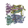

 PDBj
PDBj


 Assembly
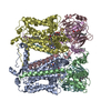


 Mass: 616.487 Da / Num. of mol.: 4 / Source method: obtained synthetically / Formula: C34H32FeN4O4
 Mass: 514.650 Da / Num. of mol.: 2 / Source method: obtained synthetically / Formula: C30H42O7
 Mass: 618.503 Da / Num. of mol.: 2 / Source method: obtained synthetically / Formula: C34H34FeN4O4
 Mass: 175.820 Da / Num. of mol.: 2 / Source method: obtained synthetically / Formula: Fe2S2
 Sample preparation
 / Beamline: I03 / Wavelength: 0.9763
  Processing