 Movie
Movie Controller


+ Open data

- Basic information
| Entry | Database: PDB / ID: 2yij | ||||||
|---|---|---|---|---|---|---|---|
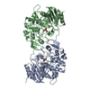
| Title | Crystal Structure of phospholipase A1 | ||||||
 Components Components | PHOSPHOLIPASE A1-IIGAMMA | ||||||
 Keywords Keywords |  HYDROLASE / PHOSPHOLIPASE HYDROLASE / PHOSPHOLIPASE | ||||||
| Function / homology |  Function and homology information Function and homology informationdiacylglycerol catabolic process / negative regulation of seed germination / monoacylglycerol catabolic process / phospholipase A1 activity / acylglycerol lipase activity / lipid storage / Hydrolases; Acting on ester bonds; Carboxylic-ester hydrolases / cytoplasmSimilarity search - Function | ||||||
| Biological species |  ARABIDOPSIS THALIANA (thale cress) ARABIDOPSIS THALIANA (thale cress) | ||||||
| Method | X-RAY DIFFRACTION / SYNCHROTRON / MAD / Resolution: 2 Å | ||||||
 Authors Authors | Lee, I. | ||||||
 Citation Citation | Journal: To be Published Title: Crystal Structure of Phospholipase A1 Authors: Lee, I. | ||||||
| History |
|
- Structure visualization
| Structure viewer | Molecule: MolmilJmol/JSmol |
|---|
- Downloads & links
-Download
| PDBx/mmCIF format | 2yij.cif.gz | 164.5 KB | Display | PDBx/mmCIF format |
|---|---|---|---|---|
| PDB format | pdb2yij.ent.gz | 138.2 KB | Display | PDB format |
| PDBx/mmJSON format | 2yij.json.gz | Tree view | PDBx/mmJSON format | |
| Others |  Other downloads Other downloads |
-Validation report
| Arichive directory | https://data.pdbj.org/pub/pdb/validation_reports/yi/2yijftp://data.pdbj.org/pub/pdb/validation_reports/yi/2yij | HTTPS FTP |
|---|
-Related structure data
| Similar structure data |
|---|


-Links
 PDBj
PDBj- Assembly
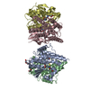
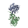
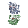
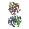
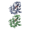
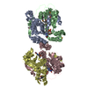
| Deposited unit | 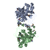
| ||||||||
|---|---|---|---|---|---|---|---|---|---|
| 1 |
| ||||||||
| Unit cell |
|
-Components
| #1: Protein | Mass: 48229.980 Da / Num. of mol.: 2 Source method: isolated from a genetically manipulated source Source: (gene. exp.) ARABIDOPSIS THALIANA (thale cress) / Production host:  ESCHERICHIA COLI (E. coli) / Strain (production host): B834(DE3) ESCHERICHIA COLI (E. coli) / Strain (production host): B834(DE3)References: UniProt: O49523, Hydrolases; Acting on ester bonds; Carboxylic-ester hydrolases#2: Water | ChemComp-HOH / | Water Mass: 18.015 Da / Num. of mol.: 406 / Source method: isolated from a natural source / Formula: H2O Mass: 18.015 Da / Num. of mol.: 406 / Source method: isolated from a natural source / Formula: H2O |
|---|
-Experimental details
-Experiment
| Experiment | Method: X-RAY DIFFRACTION |
|---|
- Sample preparation
| Crystal | Density Matthews: 2.77 Å3/Da / Density % sol: 55.57 % / Description: NONE |
|---|
-Data collection
| Diffraction | Mean temperature: 93 K | ||||||||||||
|---|---|---|---|---|---|---|---|---|---|---|---|---|---|
| Diffraction source | Source: SYNCHROTRON / Site: PAL/PLS  / Beamline: 4A / Wavelength: 0.979632, 0.979454, 0.971679 / Beamline: 4A / Wavelength: 0.979632, 0.979454, 0.971679 | ||||||||||||
| Detector | Type: ADSC CCD / Detector: CCD | ||||||||||||
| Radiation | Protocol: MAD / Monochromatic (M) / Laue (L): M / Scattering type: x-ray | ||||||||||||
| Radiation wavelength |
| ||||||||||||
| Reflection | Resolution: 1.7→30 Å / Num. obs: 114783 / % possible obs: 95.6 % / Observed criterion σ(I): 0 / Redundancy: 4.4 % / Rmerge(I) obs: 0.11 / Net I/σ(I): 25.39 | ||||||||||||
| Reflection shell | Resolution: 1.7→1.73 Å / Redundancy: 8.2 % / Rmerge(I) obs: 0.27 / Mean I/σ(I) obs: 3.45 / % possible all: 91.8 |
- Processing
| Software |
| ||||||||||||||||||||
|---|---|---|---|---|---|---|---|---|---|---|---|---|---|---|---|---|---|---|---|---|---|
| Refinement | Method to determine structure: MAD Starting model: NONE Resolution: 2→19.92 Å / Cor.coef. Fo:Fc: 0.912 / Cor.coef. Fo:Fc free: 0.887 / SU B: 0.003 / SU ML: 0 / Cross valid method: THROUGHOUT / ESU R: 3.999 / ESU R Free: 4.568 / Stereochemistry target values: MAXIMUM LIKELIHOOD / Details: HYDROGENS HAVE BEEN ADDED IN THE RIDING POSITIONS.
| ||||||||||||||||||||
| Solvent computation | Ion probe radii: 0.8 Å / Shrinkage radii: 0.8 Å / VDW probe radii: 1.4 Å / Solvent model: MASK | ||||||||||||||||||||
| Displacement parameters | Biso mean: 21.831 Å2
| ||||||||||||||||||||
| Refinement step | Cycle: LAST / Resolution: 2→19.92 Å
| ||||||||||||||||||||
| LS refinement shell | Resolution: 2→2.051 Å / Total num. of bins used: 20
|
