 Movie
Movie Controller

[English] 日本語
 Yorodumi
- PDB-2y56: Fragment growing induces conformational changes in acetylcholine-... -
+ Open data

- Basic information
| Entry | Database: PDB / ID: 2y56 | ||||||
|---|---|---|---|---|---|---|---|
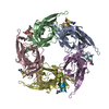
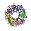
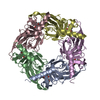
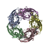
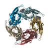
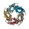
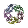
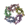
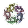
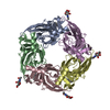
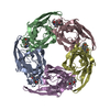
| Title | Fragment growing induces conformational changes in acetylcholine- binding protein: A structural and thermodynamic analysis - (Compound 3) | ||||||
 Components Components | SOLUBLE ACETYLCHOLINE RECEPTOR | ||||||
 Keywords Keywords |  RECEPTOR RECEPTOR | ||||||
| Function / homology |  Function and homology information Function and homology informationextracellular ligand-gated monoatomic ion channel activity / transmembrane signaling receptor activity / membrane / identical protein binding / metal ion bindingSimilarity search - Function | ||||||
| Biological species |  APLYSIA CALIFORNICA (California sea hare) APLYSIA CALIFORNICA (California sea hare) | ||||||
| Method | X-RAY DIFFRACTION / SYNCHROTRON / MOLECULAR REPLACEMENT / Resolution: 3.59 Å | ||||||
 Authors Authors | Rucktooa, P. / Edink, E. / deEsch, I.J.P. / Sixma, T.K. | ||||||
 Citation Citation | Journal: J.Am.Chem.Soc. / Year: 2011 Title: Fragment Growing Induces Conformational Changes in Acetylcholine-Binding Protein: A Structural and Thermodynamic Analysis. Authors: Edink, E. / Rucktooa, P. / Retra, K. / Akdemir, A. / Nahar, T. / Zuiderveld, O. / Van Elk, R. / Janssen, E. / Van Nierop, P. / Van Muijlwijk-Koezen, J. / Smit, A.B. / Sixma, T.K. / Leurs, R. / De Esch, I.J.P. | ||||||
| History |
|
- Structure visualization
| Structure viewer | Molecule: MolmilJmol/JSmol |
|---|
- Downloads & links
-Download
| PDBx/mmCIF format | 2y56.cif.gz | 426.9 KB | Display | PDBx/mmCIF format |
|---|---|---|---|---|
| PDB format | pdb2y56.ent.gz | 358.7 KB | Display | PDB format |
| PDBx/mmJSON format | 2y56.json.gz | Tree view | PDBx/mmJSON format | |
| Others |  Other downloads Other downloads |
-Validation report
| Arichive directory | https://data.pdbj.org/pub/pdb/validation_reports/y5/2y56ftp://data.pdbj.org/pub/pdb/validation_reports/y5/2y56 | HTTPS FTP |
|---|
-Related structure data
| Related structure data |  2y54C  2y57C  2y58C  2c9tS S: Starting model for refinement C: citing same article ( |
|---|---|
| Similar structure data |


-Links
 PDBj
PDBj

- Assembly
| Deposited unit | 
| ||||||||
|---|---|---|---|---|---|---|---|---|---|
| 1 |
| ||||||||
| Unit cell |
|
-Components
| #1: Protein | Mass: 24688.578 Da / Num. of mol.: 5 / Fragment: RESIDUES 20-236 Source method: isolated from a genetically manipulated source Source: (gene. exp.) APLYSIA CALIFORNICA (California sea hare)Plasmid: PFASTBAC1 / Cell line (production host): Sf21 / Production host:   SPODOPTERA FRUGIPERDA (fall armyworm) / References: UniProt: Q8WSF8 SPODOPTERA FRUGIPERDA (fall armyworm) / References: UniProt: Q8WSF8#2: Chemical | ChemComp-V11 / [(   Mass: 351.439 Da / Num. of mol.: 5 / Source method: obtained synthetically / Formula: C22H25NO3 Mass: 351.439 Da / Num. of mol.: 5 / Source method: obtained synthetically / Formula: C22H25NO3#3: Chemical | ChemComp-CL / Chloride  Mass: 35.453 Da / Num. of mol.: 12 / Source method: obtained synthetically / Formula: Cl Mass: 35.453 Da / Num. of mol.: 12 / Source method: obtained synthetically / Formula: Cl#4: Chemical | ChemComp-SO4 / Sulfate  Mass: 96.063 Da / Num. of mol.: 14 / Source method: obtained synthetically / Formula: SO4 Mass: 96.063 Da / Num. of mol.: 14 / Source method: obtained synthetically / Formula: SO4#5: Chemical | ChemComp-GOL / Glycerol  Mass: 92.094 Da / Num. of mol.: 8 / Source method: obtained synthetically / Formula: C3H8O3 Mass: 92.094 Da / Num. of mol.: 8 / Source method: obtained synthetically / Formula: C3H8O3 |
|---|
-Experimental details
-Experiment
| Experiment | Method: X-RAY DIFFRACTION / Number of used crystals: 1 |
|---|
- Sample preparation
| Crystal | Density Matthews: 3.77 Å3/Da / Density % sol: 67.36 % / Description: NONE |
|---|---|
| Crystal grow | pH: 8.5 / Details: 0.1M MMT PH8.5, 1.1M AMMONIUM SULPHATE |
-Data collection
| Diffraction | Mean temperature: 100 K |
|---|---|
| Diffraction source | Source: SYNCHROTRON / Site: SLS  / Beamline: X06SA / Wavelength: 1.282 / Beamline: X06SA / Wavelength: 1.282 |
| Detector | Type: DECTRIS PILATUS 6M / Detector: PIXEL / Date: Feb 5, 2010 / Details: DYNAMICALLY BENDABLE MIRROR |
| Radiation | Protocol: SINGLE WAVELENGTH / Monochromatic (M) / Laue (L): M / Scattering type: x-ray |
| Radiation wavelength | Wavelength: 1.282 Å / Relative weight: 1 |
| Reflection | Resolution: 3.59→16.05 Å / Num. obs: 20509 / % possible obs: 99.8 % / Observed criterion σ(I): 2.87 / Redundancy: 5.41 % / Biso Wilson estimate: 61.09 Å2 / Rmerge(I) obs: 0.14 / Net I/σ(I): 12.31 |
| Reflection shell | Resolution: 3.59→3.68 Å / Redundancy: 5 % / Rmerge(I) obs: 0.6 / Mean I/σ(I) obs: 2.87 / % possible all: 99.7 |
- Processing
| Software |
| ||||||||||||||||||||||||||||||||||||||||||||||||||||||||||||||||||||||||||||||||||||||||||||||||||||||||||||||||||||||||||||||||||||||||||||||||||||||
|---|---|---|---|---|---|---|---|---|---|---|---|---|---|---|---|---|---|---|---|---|---|---|---|---|---|---|---|---|---|---|---|---|---|---|---|---|---|---|---|---|---|---|---|---|---|---|---|---|---|---|---|---|---|---|---|---|---|---|---|---|---|---|---|---|---|---|---|---|---|---|---|---|---|---|---|---|---|---|---|---|---|---|---|---|---|---|---|---|---|---|---|---|---|---|---|---|---|---|---|---|---|---|---|---|---|---|---|---|---|---|---|---|---|---|---|---|---|---|---|---|---|---|---|---|---|---|---|---|---|---|---|---|---|---|---|---|---|---|---|---|---|---|---|---|---|---|---|---|---|---|---|
| Refinement | Method to determine structure: MOLECULAR REPLACEMENT Starting model: PDB ENTRY 2C9T Resolution: 3.59→48.95 Å / Cor.coef. Fo:Fc: 0.9385 / Cor.coef. Fo:Fc free: 0.9204 / Cross valid method: THROUGHOUT / σ(F): 0
| ||||||||||||||||||||||||||||||||||||||||||||||||||||||||||||||||||||||||||||||||||||||||||||||||||||||||||||||||||||||||||||||||||||||||||||||||||||||
| Displacement parameters | Biso mean: 98.43 Å2
| ||||||||||||||||||||||||||||||||||||||||||||||||||||||||||||||||||||||||||||||||||||||||||||||||||||||||||||||||||||||||||||||||||||||||||||||||||||||
| Refine analyze | Luzzati coordinate error obs: 0.662 Å | ||||||||||||||||||||||||||||||||||||||||||||||||||||||||||||||||||||||||||||||||||||||||||||||||||||||||||||||||||||||||||||||||||||||||||||||||||||||
| Refinement step | Cycle: LAST / Resolution: 3.59→48.95 Å
| ||||||||||||||||||||||||||||||||||||||||||||||||||||||||||||||||||||||||||||||||||||||||||||||||||||||||||||||||||||||||||||||||||||||||||||||||||||||
| Refine LS restraints |
| ||||||||||||||||||||||||||||||||||||||||||||||||||||||||||||||||||||||||||||||||||||||||||||||||||||||||||||||||||||||||||||||||||||||||||||||||||||||
| LS refinement shell | Resolution: 3.59→3.78 Å / Total num. of bins used: 10
| ||||||||||||||||||||||||||||||||||||||||||||||||||||||||||||||||||||||||||||||||||||||||||||||||||||||||||||||||||||||||||||||||||||||||||||||||||||||
| Refinement TLS params. | Method: refined / Refine-ID: X-RAY DIFFRACTION
| ||||||||||||||||||||||||||||||||||||||||||||||||||||||||||||||||||||||||||||||||||||||||||||||||||||||||||||||||||||||||||||||||||||||||||||||||||||||
| Refinement TLS group |
|
