 Movie
Movie Controller


+ Open data

- Basic information
| Entry | Database: PDB / ID: 2y23 | ||||||
|---|---|---|---|---|---|---|---|
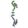
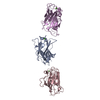
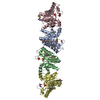
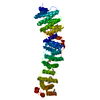
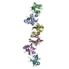
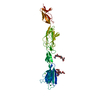
| Title | CRYSTAL STRUCTURE OF THE MYOMESIN DOMAINS MY9-MY11 | ||||||
 Components Components | MYOMESIN | ||||||
 Keywords Keywords | STRUCTURAL PROTEIN / SARCOMERE / M-BAND / IMMUNOGLOBULIN- LIKE DOMAIN | ||||||
| Function / homology |  Function and homology information Function and homology informationextraocular skeletal muscle development / striated muscle myosin thick filament / M band / structural constituent of muscle / protein kinase A signaling / positive regulation of protein secretion / kinase binding / positive regulation of gene expression / protein homodimerization activity / identical protein bindingSimilarity search - Function | ||||||
| Biological species |  HOMO SAPIENS (human) HOMO SAPIENS (human) | ||||||
| Method | X-RAY DIFFRACTION / SYNCHROTRON / MOLECULAR REPLACEMENT / Resolution: 2.5 Å | ||||||
 Authors Authors | Pinotsis, N. / Chatziefthimiou, S.D. / Wilmanns, M. | ||||||
 Citation Citation | Journal: Plos Biol. / Year: 2012 Title: Superhelical Architecture of the Myosin Filament-Linking Protein Myomesin with Unusual Elastic Properties. Authors: Pinotsis, N. / Chatziefthimiou, S.D. / Berkemeier, F. / Beuron, F. / Mavridis, I.M. / Konarev, P.V. / Svergun, D.I. / Morris, E. / Rief, M. / Wilmanns, M. | ||||||
| History |
|
- Structure visualization
| Structure viewer | Molecule: MolmilJmol/JSmol |
|---|
- Downloads & links
-Download
| PDBx/mmCIF format | 2y23.cif.gz | 142.8 KB | Display | PDBx/mmCIF format |
|---|---|---|---|---|
| PDB format | pdb2y23.ent.gz | 111.7 KB | Display | PDB format |
| PDBx/mmJSON format | 2y23.json.gz | Tree view | PDBx/mmJSON format | |
| Others |  Other downloads Other downloads |
-Validation report
| Arichive directory | https://data.pdbj.org/pub/pdb/validation_reports/y2/2y23ftp://data.pdbj.org/pub/pdb/validation_reports/y2/2y23 | HTTPS FTP |
|---|
-Related structure data
| Related structure data |  2y25C 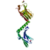 3rbsC  2r15S C: citing same article ( S: Starting model for refinement |
|---|---|
| Similar structure data |


-Links
 PDBj
PDBj


- Assembly
| Deposited unit | 
| ||||||||
|---|---|---|---|---|---|---|---|---|---|
| 1 |
| ||||||||
| Unit cell |
|
-Components
| #1: Protein | / 190 KDA CONNECTIN-ASSOCIATED PROTEIN / 190 KDA TITIN-ASSOCIATED PROTEIN / MYOMESIN FAMILY MEMBER 1 Mass: 36045.805 Da / Num. of mol.: 1 / Fragment: DOMAINS MY9, MY10, MY11, RESIDUES 1141-1447 Source method: isolated from a genetically manipulated source Source: (gene. exp.) HOMO SAPIENS (human) / Tissue: MUSCLESkeletal muscle / Production host:  ESCHERICHIA COLI (E. coli) / Strain (production host): BL21(DE3) / References: UniProt: P52179 ESCHERICHIA COLI (E. coli) / Strain (production host): BL21(DE3) / References: UniProt: P52179 | ||||
|---|---|---|---|---|---|
| #2: Chemical | 2-Mercaptoethanol  Mass: 78.133 Da / Num. of mol.: 3 / Source method: obtained synthetically / Formula: C2H6OS Mass: 78.133 Da / Num. of mol.: 3 / Source method: obtained synthetically / Formula: C2H6OS#3: Chemical | ChemComp-PG0 / | 2-(2-Methoxyethoxy)ethanol  Mass: 120.147 Da / Num. of mol.: 1 / Source method: obtained synthetically / Formula: C5H12O3 / Comment: inhibitor, precipitant*YM Mass: 120.147 Da / Num. of mol.: 1 / Source method: obtained synthetically / Formula: C5H12O3 / Comment: inhibitor, precipitant*YM#4: Water | ChemComp-HOH / | Water Mass: 18.015 Da / Num. of mol.: 182 / Source method: isolated from a natural source / Formula: H2O Mass: 18.015 Da / Num. of mol.: 182 / Source method: isolated from a natural source / Formula: H2O |
-Experimental details
-Experiment
| Experiment | Method: X-RAY DIFFRACTION / Number of used crystals: 1 |
|---|
- Sample preparation
| Crystal | Density Matthews: 3.11 Å3/Da / Density % sol: 60.5 % / Description: NONE |
|---|---|
| Crystal grow | pH: 7.5 Details: 0.18M MAGNESIUM ACETATE 20% W/V POLYETHYLENE GLYCOL 3350, pH 7.5 |
-Data collection
| Diffraction | Mean temperature: 100 K |
|---|---|
| Diffraction source | Source: SYNCHROTRON / Site: EMBL/DESY, HAMBURG  / Beamline: X12 / Wavelength: 0.97699 / Beamline: X12 / Wavelength: 0.97699 |
| Detector | Type: MARRESEARCH / Detector: CCD / Details: MIRRORS |
| Radiation | Monochromator: DOUBLE CRYSTAL SI(111), HORIZONTALLY FOCUSSING Protocol: SINGLE WAVELENGTH / Monochromatic (M) / Laue (L): M / Scattering type: x-ray |
| Radiation wavelength | Wavelength: 0.97699 Å / Relative weight: 1 |
| Reflection | Resolution: 2.49→25 Å / Num. obs: 15174 / % possible obs: 99 % / Observed criterion σ(I): -3 / Redundancy: 3.3 % / Biso Wilson estimate: 58.3 Å2 / Rmerge(I) obs: 0.11 / Net I/σ(I): 10.6 |
| Reflection shell | Resolution: 2.49→2.53 Å / Redundancy: 2.9 % / Rmerge(I) obs: 0.52 / Mean I/σ(I) obs: 1.8 / % possible all: 89.6 |
- Processing
| Software |
| ||||||||||||||||||||||||||||||||||||||||||||||||||||||||||||||||||||||||||||||||||||||||||||||||||||||||||||||||||||||||||||||||||||||||||||||||||||||||||||||||||||||||||||||||||||||
|---|---|---|---|---|---|---|---|---|---|---|---|---|---|---|---|---|---|---|---|---|---|---|---|---|---|---|---|---|---|---|---|---|---|---|---|---|---|---|---|---|---|---|---|---|---|---|---|---|---|---|---|---|---|---|---|---|---|---|---|---|---|---|---|---|---|---|---|---|---|---|---|---|---|---|---|---|---|---|---|---|---|---|---|---|---|---|---|---|---|---|---|---|---|---|---|---|---|---|---|---|---|---|---|---|---|---|---|---|---|---|---|---|---|---|---|---|---|---|---|---|---|---|---|---|---|---|---|---|---|---|---|---|---|---|---|---|---|---|---|---|---|---|---|---|---|---|---|---|---|---|---|---|---|---|---|---|---|---|---|---|---|---|---|---|---|---|---|---|---|---|---|---|---|---|---|---|---|---|---|---|---|---|---|
| Refinement | Method to determine structure: MOLECULAR REPLACEMENT Starting model: MYOMESIN DOMAINS MY10-MY11, PDB ENTRY 2R15 Resolution: 2.5→25 Å / Cor.coef. Fo:Fc: 0.948 / Cor.coef. Fo:Fc free: 0.928 / SU B: 23.84 / SU ML: 0.283 / Cross valid method: THROUGHOUT / ESU R: 0.434 / ESU R Free: 0.292 / Stereochemistry target values: MAXIMUM LIKELIHOOD / Details: HYDROGENS HAVE BEEN ADDED IN THE RIDING POSITIONS.
| ||||||||||||||||||||||||||||||||||||||||||||||||||||||||||||||||||||||||||||||||||||||||||||||||||||||||||||||||||||||||||||||||||||||||||||||||||||||||||||||||||||||||||||||||||||||
| Solvent computation | Ion probe radii: 0.8 Å / Shrinkage radii: 0.8 Å / VDW probe radii: 1.2 Å / Solvent model: BABINET MODEL WITH MASK | ||||||||||||||||||||||||||||||||||||||||||||||||||||||||||||||||||||||||||||||||||||||||||||||||||||||||||||||||||||||||||||||||||||||||||||||||||||||||||||||||||||||||||||||||||||||
| Displacement parameters | Biso mean: 59.363 Å2
| ||||||||||||||||||||||||||||||||||||||||||||||||||||||||||||||||||||||||||||||||||||||||||||||||||||||||||||||||||||||||||||||||||||||||||||||||||||||||||||||||||||||||||||||||||||||
| Refinement step | Cycle: LAST / Resolution: 2.5→25 Å
| ||||||||||||||||||||||||||||||||||||||||||||||||||||||||||||||||||||||||||||||||||||||||||||||||||||||||||||||||||||||||||||||||||||||||||||||||||||||||||||||||||||||||||||||||||||||
| Refine LS restraints |
|