 Movie
Movie Controller


+ Open data

- Basic information
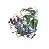
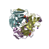
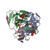
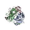
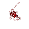
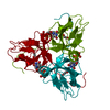
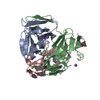
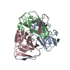
| Entry | Database: PDB / ID: 2y1t | ||||||
|---|---|---|---|---|---|---|---|
| Title | Bacillus subtilis prophage dUTPase in complex with dUDP | ||||||
 Components Components | SPBC2 PROPHAGE-DERIVED DEOXYURIDINE 5'-TRIPHOSPHATE NUCLEOTIDOHYDROLASE YOSS | ||||||
 Keywords Keywords |  HYDROLASE / SPB PROPHAGE / PHE-LID HYDROLASE / SPB PROPHAGE / PHE-LID | ||||||
| Function / homology |  Function and homology information Function and homology informationdUTP catabolic process / dUMP biosynthetic process / dUTP diphosphatase / dUTP diphosphatase activity / magnesium ion binding / protein-containing complex / identical protein bindingSimilarity search - Function | ||||||
| Biological species |  BACILLUS SUBTILIS (bacteria) BACILLUS SUBTILIS (bacteria) | ||||||
| Method | X-RAY DIFFRACTION / SYNCHROTRON / MOLECULAR REPLACEMENT / Resolution: 1.89 Å | ||||||
 Authors Authors | Garcia-Nafria, J. / Harkiolaki, M. / Persson, R. / Fogg, M.J. / Wilson, K.S. | ||||||
 Citation Citation | Journal: Acta Crystallogr.,Sect.D / Year: 2011 Title: The Structure of Bacillus Subtilis Sp Beta Prophage Dutpase and its Complexes with Two Nucleotides Authors: Garcia-Nafria, J. / Harkiolaki, M. / Persson, R. / Fogg, M.J. / Wilson, K.S. | ||||||
| History |
|
- Structure visualization
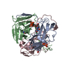
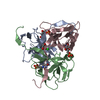
| Structure viewer | Molecule: MolmilJmol/JSmol |
|---|
- Downloads & links
-Download
| PDBx/mmCIF format | 2y1t.cif.gz | 180.8 KB | Display | PDBx/mmCIF format |
|---|---|---|---|---|
| PDB format | pdb2y1t.ent.gz | 144.7 KB | Display | PDB format |
| PDBx/mmJSON format | 2y1t.json.gz | Tree view | PDBx/mmJSON format | |
| Others |  Other downloads Other downloads |
-Validation report
| Arichive directory | https://data.pdbj.org/pub/pdb/validation_reports/y1/2y1tftp://data.pdbj.org/pub/pdb/validation_reports/y1/2y1t | HTTPS FTP |
|---|
-Related structure data
| Related structure data |  2xx6SC  2xy3C C: citing same article ( S: Starting model for refinement |
|---|---|
| Similar structure data |

-Links
 PDBj
PDBj
- Assembly
| Deposited unit | 
| ||||||||
|---|---|---|---|---|---|---|---|---|---|
| 1 | 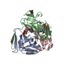
| ||||||||
| 2 | 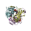
| ||||||||
| Unit cell |
|
-Components
| #1: Protein | Mass: 16172.509 Da / Num. of mol.: 6 Source method: isolated from a genetically manipulated source Source: (gene. exp.) BACILLUS SUBTILIS (bacteria) / Production host: ESCHERICHIA COLI (E. coli) / Strain (production host): B834 / References: UniProt: O34919, dUTP diphosphatase#2: Chemical | ChemComp-DUD / 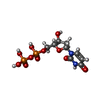  Mass: 388.162 Da / Num. of mol.: 5 / Source method: obtained synthetically / Formula: C9H14N2O11P2 Mass: 388.162 Da / Num. of mol.: 5 / Source method: obtained synthetically / Formula: C9H14N2O11P2#3: Water | ChemComp-HOH / | Water Mass: 18.015 Da / Num. of mol.: 507 / Source method: isolated from a natural source / Formula: H2O Mass: 18.015 Da / Num. of mol.: 507 / Source method: isolated from a natural source / Formula: H2O |
|---|
-Experimental details
-Experiment
| Experiment | Method: X-RAY DIFFRACTION / Number of used crystals: 1 |
|---|
- Sample preparation
| Crystal | Density Matthews: 2.27 Å3/Da / Density % sol: 46 % / Description: NONE |
|---|---|
| Crystal grow | pH: 7.5 Details: 0.1M HEPES BUFFER PH 7.5, 10% (V/V) PEG 8000, 8% ETHYLENE GLYCOL |
-Data collection
| Diffraction | Mean temperature: 100 K | ||||||||||||||||||||
|---|---|---|---|---|---|---|---|---|---|---|---|---|---|---|---|---|---|---|---|---|---|
| Diffraction source | Source: SYNCHROTRON / Site: ESRF  / Beamline: ID14-1 / Wavelength: 0.934 / Beamline: ID14-1 / Wavelength: 0.934 | ||||||||||||||||||||
| Detector | Type: ADSC QUANTUM 210 / Detector: CCD / Date: Jan 10, 2001 | ||||||||||||||||||||
| Radiation | Protocol: SINGLE WAVELENGTH / Monochromatic (M) / Laue (L): M / Scattering type: x-ray | ||||||||||||||||||||
| Radiation wavelength | Wavelength: 0.934 Å / Relative weight: 1 | ||||||||||||||||||||
| Reflection twin |
| ||||||||||||||||||||
| Reflection | Resolution: 1.9→20 Å / Num. obs: 78511 / % possible obs: 99.8 % / Observed criterion σ(I): 1 / Redundancy: 5.4 % / Rmerge(I) obs: 0.05 / Net I/σ(I): 25.4 | ||||||||||||||||||||
| Reflection shell | Resolution: 1.9→1.97 Å / Rmerge(I) obs: 0.45 / Mean I/σ(I) obs: 3.1 / % possible all: 99 |
- Processing
| Software |
| ||||||||||||||||||||||||||||||||||||||||||||||||||||||||||||||||||||||||||||||||||||||||||||||||||||||||||||||||||||||||||||||||||||||||||||||||||||||||||||||||||||||||||||||||||||||
|---|---|---|---|---|---|---|---|---|---|---|---|---|---|---|---|---|---|---|---|---|---|---|---|---|---|---|---|---|---|---|---|---|---|---|---|---|---|---|---|---|---|---|---|---|---|---|---|---|---|---|---|---|---|---|---|---|---|---|---|---|---|---|---|---|---|---|---|---|---|---|---|---|---|---|---|---|---|---|---|---|---|---|---|---|---|---|---|---|---|---|---|---|---|---|---|---|---|---|---|---|---|---|---|---|---|---|---|---|---|---|---|---|---|---|---|---|---|---|---|---|---|---|---|---|---|---|---|---|---|---|---|---|---|---|---|---|---|---|---|---|---|---|---|---|---|---|---|---|---|---|---|---|---|---|---|---|---|---|---|---|---|---|---|---|---|---|---|---|---|---|---|---|---|---|---|---|---|---|---|---|---|---|---|
| Refinement | Method to determine structure: MOLECULAR REPLACEMENT Starting model: CHAIN A OF PDB ENTRY 2XX6 Resolution: 1.89→70.31 Å / Cor.coef. Fo:Fc: 0.972 / Cor.coef. Fo:Fc free: 0.966 / SU B: 4.374 / SU ML: 0.066 / Cross valid method: THROUGHOUT / ESU R: 0.022 / ESU R Free: 0.021 / Stereochemistry target values: MAXIMUM LIKELIHOOD / Details: HYDROGENS HAVE BEEN USED IF PRESENT IN THE INPUT
| ||||||||||||||||||||||||||||||||||||||||||||||||||||||||||||||||||||||||||||||||||||||||||||||||||||||||||||||||||||||||||||||||||||||||||||||||||||||||||||||||||||||||||||||||||||||
| Solvent computation | Ion probe radii: 0.8 Å / Shrinkage radii: 0.8 Å / VDW probe radii: 1.2 Å / Solvent model: MASK | ||||||||||||||||||||||||||||||||||||||||||||||||||||||||||||||||||||||||||||||||||||||||||||||||||||||||||||||||||||||||||||||||||||||||||||||||||||||||||||||||||||||||||||||||||||||
| Displacement parameters | Biso mean: 19.95 Å2
| ||||||||||||||||||||||||||||||||||||||||||||||||||||||||||||||||||||||||||||||||||||||||||||||||||||||||||||||||||||||||||||||||||||||||||||||||||||||||||||||||||||||||||||||||||||||
| Refinement step | Cycle: LAST / Resolution: 1.89→70.31 Å
| ||||||||||||||||||||||||||||||||||||||||||||||||||||||||||||||||||||||||||||||||||||||||||||||||||||||||||||||||||||||||||||||||||||||||||||||||||||||||||||||||||||||||||||||||||||||
| Refine LS restraints |
|