 Movie
Movie Controller


+ Open data

- Basic information
| Entry | Database: PDB / ID: 2xzd | ||||||
|---|---|---|---|---|---|---|---|
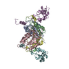
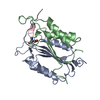
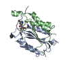
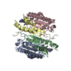
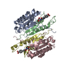
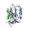
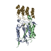
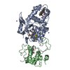
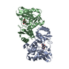
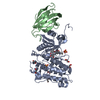
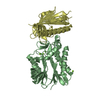
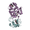
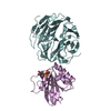
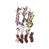
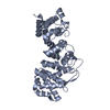
| Title | Caspase-3 in Complex with an Inhibitory DARPin-3.4 | ||||||
 Components Components |
| ||||||
 Keywords Keywords | HYDROLASE/PROTEIN BINDING / HYDROLASE-PROTEIN BINDING COMPLEX /  DE NOVO PROTEIN / APOPTOSIS / ANKYRIN REPEAT PROTEIN / RIBOSOME DISPLAY DE NOVO PROTEIN / APOPTOSIS / ANKYRIN REPEAT PROTEIN / RIBOSOME DISPLAY | ||||||
| Function / homology |  Function and homology informationcaspase-3 / Stimulation of the cell death response by PAK-2p34 / phospholipase A2 activator activity / anterior neural tube closure / intrinsic apoptotic signaling pathway in response to osmotic stress / leukocyte apoptotic process / positive regulation of pyroptosis / glial cell apoptotic process / NADE modulates death signalling / response to cobalt ion ...caspase-3 / Stimulation of the cell death response by PAK-2p34 / phospholipase A2 activator activity / anterior neural tube closure / intrinsic apoptotic signaling pathway in response to osmotic stress / leukocyte apoptotic process / positive regulation of pyroptosis / glial cell apoptotic process / NADE modulates death signalling / response to cobalt ion / cysteine-type endopeptidase activity involved in apoptotic signaling pathway / luteolysis / death-inducing signaling complex / cyclin-dependent protein serine/threonine kinase inhibitor activity / cellular response to staurosporine / Apoptosis induced DNA fragmentation / Apoptotic cleavage of cell adhesion proteins / cysteine-type endopeptidase activity involved in execution phase of apoptosis / Caspase activation via Dependence Receptors in the absence of ligand / death receptor binding / SMAC, XIAP-regulated apoptotic response / axonal fasciculation / Activation of caspases through apoptosome-mediated cleavage / Signaling by Hippo / cysteine-type endopeptidase activity involved in apoptotic process / SMAC (DIABLO) binds to IAPs / SMAC(DIABLO)-mediated dissociation of IAP:caspase complexes / fibroblast apoptotic process / execution phase of apoptosis / negative regulation of cytokine production / epithelial cell apoptotic process / platelet formation / Other interleukin signaling / positive regulation of amyloid-beta formation / pyroptosis / Apoptotic cleavage of cellular proteins / negative regulation of B cell proliferation / T cell homeostasis / negative regulation of activated T cell proliferation / neurotrophin TRK receptor signaling pathway / B cell homeostasis / protein maturation / negative regulation of cell cycle / response to X-ray / Caspase-mediated cleavage of cytoskeletal proteins / regulation of macroautophagy / response to amino acid / cell fate commitment / Pyroptosis / response to tumor necrosis factor / response to glucose / response to UV / response to glucocorticoid / keratinocyte differentiation / striated muscle cell differentiation / Degradation of the extracellular matrix / intrinsic apoptotic signaling pathway / erythrocyte differentiation / response to nicotine / apoptotic signaling pathway / hippocampus development / sensory perception of sound / protein catabolic process / regulation of protein stability / response to hydrogen peroxide / neuron differentiation / protein processing / response to wounding / positive regulation of neuron apoptotic process / response to estradiol / heart development / peptidase activity / neuron apoptotic process / protease binding / response to lipopolysaccharide / response to hypoxia / aspartic-type endopeptidase activity / learning or memory / response to xenobiotic stimulus / positive regulation of apoptotic process / cysteine-type endopeptidase activity / neuronal cell body / apoptotic process / DNA damage response / protein-containing complex binding / proteolysis / nucleoplasm / nucleus / cytosol / cytoplasm Function and homology informationcaspase-3 / Stimulation of the cell death response by PAK-2p34 / phospholipase A2 activator activity / anterior neural tube closure / intrinsic apoptotic signaling pathway in response to osmotic stress / leukocyte apoptotic process / positive regulation of pyroptosis / glial cell apoptotic process / NADE modulates death signalling / response to cobalt ion ...caspase-3 / Stimulation of the cell death response by PAK-2p34 / phospholipase A2 activator activity / anterior neural tube closure / intrinsic apoptotic signaling pathway in response to osmotic stress / leukocyte apoptotic process / positive regulation of pyroptosis / glial cell apoptotic process / NADE modulates death signalling / response to cobalt ion / cysteine-type endopeptidase activity involved in apoptotic signaling pathway / luteolysis / death-inducing signaling complex / cyclin-dependent protein serine/threonine kinase inhibitor activity / cellular response to staurosporine / Apoptosis induced DNA fragmentation / Apoptotic cleavage of cell adhesion proteins / cysteine-type endopeptidase activity involved in execution phase of apoptosis / Caspase activation via Dependence Receptors in the absence of ligand / death receptor binding / SMAC, XIAP-regulated apoptotic response / axonal fasciculation / Activation of caspases through apoptosome-mediated cleavage / Signaling by Hippo / cysteine-type endopeptidase activity involved in apoptotic process / SMAC (DIABLO) binds to IAPs / SMAC(DIABLO)-mediated dissociation of IAP:caspase complexes / fibroblast apoptotic process / execution phase of apoptosis / negative regulation of cytokine production / epithelial cell apoptotic process / platelet formation / Other interleukin signaling / positive regulation of amyloid-beta formation / pyroptosis / Apoptotic cleavage of cellular proteins / negative regulation of B cell proliferation / T cell homeostasis / negative regulation of activated T cell proliferation / neurotrophin TRK receptor signaling pathway / B cell homeostasis / protein maturation / negative regulation of cell cycle / response to X-ray / Caspase-mediated cleavage of cytoskeletal proteins / regulation of macroautophagy / response to amino acid / cell fate commitment / Pyroptosis / response to tumor necrosis factor / response to glucose / response to UV / response to glucocorticoid / keratinocyte differentiation / striated muscle cell differentiation / Degradation of the extracellular matrix / intrinsic apoptotic signaling pathway / erythrocyte differentiation / response to nicotine / apoptotic signaling pathway / hippocampus development / sensory perception of sound / protein catabolic process / regulation of protein stability / response to hydrogen peroxide / neuron differentiation / protein processing / response to wounding / positive regulation of neuron apoptotic process / response to estradiol / heart development / peptidase activity / neuron apoptotic process / protease binding / response to lipopolysaccharide / response to hypoxia / aspartic-type endopeptidase activity / learning or memory / response to xenobiotic stimulus / positive regulation of apoptotic process / cysteine-type endopeptidase activity / neuronal cell body / apoptotic process / DNA damage response / protein-containing complex binding / proteolysis / nucleoplasm / nucleus / cytosol / cytoplasmSimilarity search - Function | ||||||
| Biological species |  HOMO SAPIENS (human) HOMO SAPIENS (human)SYNTHETIC CONSTRUCT (others) | ||||||
| Method | X-RAY DIFFRACTION / SYNCHROTRON / MOLECULAR REPLACEMENT / Resolution: 2.1 Å | ||||||
 Authors Authors | Barandun, J. / Schroeder, T. / Mittl, P. / Grutter, M.G. | ||||||
 Citation Citation | Journal: Structure / Year: 2013 Title: Specific Inhibition of Caspase-3 by a Competitive Darpin: Molecular Mimicry between Native and Designed Inhibitors. Authors: Schroeder, T. / Barandun, J. / Flutsch, A. / Briand, C. / Mittl, P. / Grutter, M.G. | ||||||
| History |
|
- Structure visualization
| Structure viewer | Molecule: MolmilJmol/JSmol |
|---|
- Downloads & links
-Download
| PDBx/mmCIF format | 2xzd.cif.gz | 301.2 KB | Display | PDBx/mmCIF format |
|---|---|---|---|---|
| PDB format | pdb2xzd.ent.gz | 245.8 KB | Display | PDB format |
| PDBx/mmJSON format | 2xzd.json.gz | Tree view | PDBx/mmJSON format | |
| Others |  Other downloads Other downloads |
-Validation report
| Arichive directory | https://data.pdbj.org/pub/pdb/validation_reports/xz/2xzdftp://data.pdbj.org/pub/pdb/validation_reports/xz/2xzd | HTTPS FTP |
|---|
-Related structure data
| Related structure data |  2y0bC  2dkoS S: Starting model for refinement C: citing same article ( |
|---|---|
| Similar structure data |


-Links
 PDBj
PDBj


- Assembly
| Deposited unit | 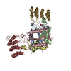
| ||||||||
|---|---|---|---|---|---|---|---|---|---|
| 1 | 
| ||||||||
| 2 | 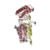
| ||||||||
| Unit cell |
|
-Components
-Protein , 3 types, 6 molecules ACBDGH
| #1: Protein | Caspase 3 / CASP-3 / APOPAIN / CYSTEINE PROTEASE CPP32 / CPP-32 PROTEIN YAM / SREBP CLEAVAGE ACTIVITY 1 / SCA-1 Mass: 16874.174 Da / Num. of mol.: 2 / Fragment: P17 SUBUNIT, RESIDUES 29-175 / Mutation: YES Source method: isolated from a genetically manipulated source Source: (gene. exp.) HOMO SAPIENS (human) / Production host:  ESCHERICHIA COLI (E. coli) / Strain (production host): BL21(DE3) / References: UniProt: P42574, caspase-3 ESCHERICHIA COLI (E. coli) / Strain (production host): BL21(DE3) / References: UniProt: P42574, caspase-3#2: Protein | Caspase 3 / CASP-3 / APOPAIN / CYSTEINE PROTEASE CPP32 / CPP-32 PROTEIN YAM / SREBP CLEAVAGE ACTIVITY 1 / SCA-1Mass: 13832.739 Da / Num. of mol.: 2 / Fragment: P12 SUBUNIT, RESIDUES 176-277 Source method: isolated from a genetically manipulated source Source: (gene. exp.) HOMO SAPIENS (human) / Production host: ESCHERICHIA COLI (E. coli) / Strain (production host): BL21(DE3) / References: UniProt: P42574, caspase-3#3: Protein | Mass: 14740.521 Da / Num. of mol.: 2 Source method: isolated from a genetically manipulated source Details: IN-VITRO EVOLVED SEQUENCE / Source: (gene. exp.) SYNTHETIC CONSTRUCT (others) / Production host: ESCHERICHIA COLI (E. coli) / Strain (production host): K-12 / Variant (production host): XL1BLUE |
|---|
-Non-polymers , 3 types, 321 molecules 


| #4: Chemical | 2-Methyl-2,4-pentanediol Mass: 118.174 Da / Num. of mol.: 3 / Source method: obtained synthetically / Formula: C6H14O2 / Comment: precipitant*YM Mass: 118.174 Da / Num. of mol.: 3 / Source method: obtained synthetically / Formula: C6H14O2 / Comment: precipitant*YM#5: Chemical | ChemComp-MPD / ( | 2-Methyl-2,4-pentanediol Mass: 118.174 Da / Num. of mol.: 1 / Source method: obtained synthetically / Formula: C6H14O2 / Comment: precipitant*YM Mass: 118.174 Da / Num. of mol.: 1 / Source method: obtained synthetically / Formula: C6H14O2 / Comment: precipitant*YM#6: Water | ChemComp-HOH / | WaterMass: 18.015 Da / Num. of mol.: 317 / Source method: isolated from a natural source / Formula: H2O |
|---|
-Details
| Sequence details | CHAIN A AND C, ASP 28 TO SER MUTATIONS FROM CLONING STRATEGY. |
|---|
-Experimental details
-Experiment
| Experiment | Method: X-RAY DIFFRACTION / Number of used crystals: 3 |
|---|
- Sample preparation
| Crystal | Density Matthews: 3.11 Å3/Da / Density % sol: 60 % / Description: NONE |
|---|---|
| Crystal grow | pH: 7.3 Details: 100 MM HEPES, PH 7.3 (RT), 40 % 2-METHYL-2,4-PENTANEDIOL. |
-Data collection
| Diffraction | Mean temperature: 90 K |
|---|---|
| Diffraction source | Source: SYNCHROTRON / Site: SLS  / Beamline: X06SA / Wavelength: 1 / Beamline: X06SA / Wavelength: 1 |
| Detector | Type: DECTRIS PILATUS 6M / Detector: PIXEL / Date: Oct 12, 2010 |
| Radiation | Monochromator: SI / Protocol: SINGLE WAVELENGTH / Monochromatic (M) / Laue (L): M / Scattering type: x-ray |
| Radiation wavelength | Wavelength: 1 Å / Relative weight: 1 |
| Reflection | Resolution: 2.1→49 Å / Num. obs: 63172 / % possible obs: 99.3 % / Observed criterion σ(I): 1.92 / Redundancy: 3.9 % / Biso Wilson estimate: 35.29 Å2 / Rmerge(I) obs: 0.04 / Net I/σ(I): 15.59 |
| Reflection shell | Resolution: 2.1→2.2 Å / Redundancy: 4.03 % / Rmerge(I) obs: 0.6 / Mean I/σ(I) obs: 1.92 / % possible all: 99.5 |
- Processing
| Software |
| ||||||||||||||||||||||||||||||||||||||||||||||||||||||||||||||||||||||||||||||||||||||||||||||||||||||||||||||||||||||||||||||||||||||||||||||||||||||||||||||||||||||||||||||||||||||
|---|---|---|---|---|---|---|---|---|---|---|---|---|---|---|---|---|---|---|---|---|---|---|---|---|---|---|---|---|---|---|---|---|---|---|---|---|---|---|---|---|---|---|---|---|---|---|---|---|---|---|---|---|---|---|---|---|---|---|---|---|---|---|---|---|---|---|---|---|---|---|---|---|---|---|---|---|---|---|---|---|---|---|---|---|---|---|---|---|---|---|---|---|---|---|---|---|---|---|---|---|---|---|---|---|---|---|---|---|---|---|---|---|---|---|---|---|---|---|---|---|---|---|---|---|---|---|---|---|---|---|---|---|---|---|---|---|---|---|---|---|---|---|---|---|---|---|---|---|---|---|---|---|---|---|---|---|---|---|---|---|---|---|---|---|---|---|---|---|---|---|---|---|---|---|---|---|---|---|---|---|---|---|---|
| Refinement | Method to determine structure: MOLECULAR REPLACEMENT Starting model: PDB ENTRY 2DKO Resolution: 2.1→49 Å / Cor.coef. Fo:Fc: 0.97 / Cor.coef. Fo:Fc free: 0.959 / SU B: 10.66 / SU ML: 0.119 / Cross valid method: THROUGHOUT / ESU R: 0.161 / ESU R Free: 0.147 / Stereochemistry target values: MAXIMUM LIKELIHOOD / Details: HYDROGENS HAVE BEEN ADDED IN THE RIDING POSITIONS.
| ||||||||||||||||||||||||||||||||||||||||||||||||||||||||||||||||||||||||||||||||||||||||||||||||||||||||||||||||||||||||||||||||||||||||||||||||||||||||||||||||||||||||||||||||||||||
| Solvent computation | Ion probe radii: 0.8 Å / Shrinkage radii: 0.8 Å / VDW probe radii: 1.4 Å / Solvent model: MASK | ||||||||||||||||||||||||||||||||||||||||||||||||||||||||||||||||||||||||||||||||||||||||||||||||||||||||||||||||||||||||||||||||||||||||||||||||||||||||||||||||||||||||||||||||||||||
| Displacement parameters | Biso mean: 55.8 Å2
| ||||||||||||||||||||||||||||||||||||||||||||||||||||||||||||||||||||||||||||||||||||||||||||||||||||||||||||||||||||||||||||||||||||||||||||||||||||||||||||||||||||||||||||||||||||||
| Refinement step | Cycle: LAST / Resolution: 2.1→49 Å
| ||||||||||||||||||||||||||||||||||||||||||||||||||||||||||||||||||||||||||||||||||||||||||||||||||||||||||||||||||||||||||||||||||||||||||||||||||||||||||||||||||||||||||||||||||||||
| Refine LS restraints |
|