 Movie
Movie Controller


+ Open data

- Basic information
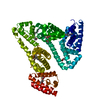
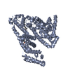
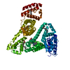
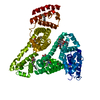
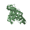
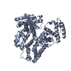
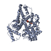
| Entry | Database: PDB / ID: 2xvu | ||||||
|---|---|---|---|---|---|---|---|
| Title | Human serum albumin complexed with dansyl-L-asparagine | ||||||
 Components Components | SERUM ALBUMIN | ||||||
 Keywords Keywords | TRANSPORT PROTEIN | ||||||
| Function / homology |  Function and homology information Function and homology informationcellular response to calcium ion starvation / exogenous protein binding / Ciprofloxacin ADME / HDL remodeling / enterobactin binding / Heme biosynthesis / negative regulation of mitochondrial depolarization / Prednisone ADME / Heme degradation / antioxidant activity ...cellular response to calcium ion starvation / exogenous protein binding / Ciprofloxacin ADME / HDL remodeling / enterobactin binding / Heme biosynthesis / negative regulation of mitochondrial depolarization / Prednisone ADME / Heme degradation / antioxidant activity / Aspirin ADME / toxic substance binding / Scavenging of heme from plasma / Recycling of bile acids and salts / cellular response to starvation / platelet alpha granule lumen / fatty acid binding / Post-translational protein phosphorylation / Cytoprotection by HMOX1 / Regulation of Insulin-like Growth Factor (IGF) transport and uptake by Insulin-like Growth Factor Binding Proteins (IGFBPs) / pyridoxal phosphate binding / Platelet degranulation / protein-folding chaperone binding / blood microparticle / copper ion binding / endoplasmic reticulum lumen / Golgi apparatus / endoplasmic reticulum / protein-containing complex / DNA binding / extracellular space / extracellular exosome / extracellular region / identical protein binding / nucleus / cytoplasmSimilarity search - Function | ||||||
| Biological species |  HOMO SAPIENS (human) HOMO SAPIENS (human) | ||||||
| Method | X-RAY DIFFRACTION / SYNCHROTRON / MOLECULAR REPLACEMENT / Resolution: 2.6 Å | ||||||
 Authors Authors | Zunszain, P.A. / Curry, S. | ||||||
 Citation Citation | Journal: J.Struct.Biol. / Year: 2011 Title: Structural Basis of Binding of Fluorescent, Site-Specific Dansylated Amino Acids to Human Serum Albumin. Authors: Ryan, A.J. / Ghuman, J. / Zunszain, P.A. / Chung, C.W. / Curry, S. | ||||||
| History |
|
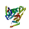
- Structure visualization
| Structure viewer | Molecule: MolmilJmol/JSmol |
|---|
- Downloads & links
-Download
| PDBx/mmCIF format | 2xvu.cif.gz | 220.9 KB | Display | PDBx/mmCIF format |
|---|---|---|---|---|
| PDB format | pdb2xvu.ent.gz | 174.4 KB | Display | PDB format |
| PDBx/mmJSON format | 2xvu.json.gz | Tree view | PDBx/mmJSON format | |
| Others |  Other downloads Other downloads |
-Validation report
| Arichive directory | https://data.pdbj.org/pub/pdb/validation_reports/xv/2xvuftp://data.pdbj.org/pub/pdb/validation_reports/xv/2xvu | HTTPS FTP |
|---|
-Related structure data
| Related structure data |  2xsiC  2xvqC  2xvvC  2xvwC  2xw0C  2xw1C  2bxaS C: citing same article ( S: Starting model for refinement |
|---|---|
| Similar structure data |


-Links
 PDBj
PDBj


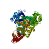
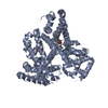
- Assembly
| Deposited unit | 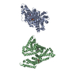
| ||||||||
|---|---|---|---|---|---|---|---|---|---|
| 1 | 
| ||||||||
| 2 | 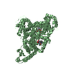
| ||||||||
| Unit cell |
|
-Components
| #1: Protein | Mass: 66571.219 Da / Num. of mol.: 2 Source method: isolated from a genetically manipulated source Source: (gene. exp.) HOMO SAPIENS (human) / Production host:  PICHIA PASTORIS (fungus) / References: UniProt: P02768 PICHIA PASTORIS (fungus) / References: UniProt: P02768#2: Chemical | ChemComp-9DN /   Type: L-peptide linking / Mass: 365.404 Da / Num. of mol.: 4 / Source method: obtained synthetically / Formula: C16H19N3O5S Type: L-peptide linking / Mass: 365.404 Da / Num. of mol.: 4 / Source method: obtained synthetically / Formula: C16H19N3O5S#3: Water | ChemComp-HOH / | Water Mass: 18.015 Da / Num. of mol.: 35 / Source method: isolated from a natural source / Formula: H2O Mass: 18.015 Da / Num. of mol.: 35 / Source method: isolated from a natural source / Formula: H2OSequence details | SEQUENCE NUMBERING IS OF MATURE (SECRETED) PROTEIN. | |
|---|
-Experimental details
-Experiment
| Experiment | Method: X-RAY DIFFRACTION / Number of used crystals: 1 |
|---|
- Sample preparation
| Crystal | Density Matthews: 2.6 Å3/Da / Density % sol: 53 % / Description: NONE |
|---|---|
| Crystal grow | pH: 7 / Details: SEE PAPER, pH 7 |
-Data collection
| Diffraction | Mean temperature: 298 K |
|---|---|
| Diffraction source | Source: SYNCHROTRON / Site: SRS  / Beamline: PX9.6 / Wavelength: 0.87 / Beamline: PX9.6 / Wavelength: 0.87 |
| Detector | Type: ADSC QUANTUM 4 / Detector: CCD / Date: Mar 21, 2007 / Details: MIRRORS |
| Radiation | Monochromator: CRYSTAL / Protocol: SINGLE WAVELENGTH / Monochromatic (M) / Laue (L): M / Scattering type: x-ray |
| Radiation wavelength | Wavelength: 0.87 Å / Relative weight: 1 |
| Reflection | Resolution: 2.6→35.1 Å / Num. obs: 37571 / % possible obs: 97.7 % / Observed criterion σ(I): 0 / Redundancy: 2 % / Biso Wilson estimate: 42.7 Å2 / Rmerge(I) obs: 0.05 / Net I/σ(I): 10.4 |
| Reflection shell | Resolution: 2.6→2.76 Å / Redundancy: 2 % / Rmerge(I) obs: 0.38 / Mean I/σ(I) obs: 2 / % possible all: 97.4 |
- Processing
| Software |
| ||||||||||||||||||||||||||||||||||||||||||||||||||||||||||||||||||||||||||||||||
|---|---|---|---|---|---|---|---|---|---|---|---|---|---|---|---|---|---|---|---|---|---|---|---|---|---|---|---|---|---|---|---|---|---|---|---|---|---|---|---|---|---|---|---|---|---|---|---|---|---|---|---|---|---|---|---|---|---|---|---|---|---|---|---|---|---|---|---|---|---|---|---|---|---|---|---|---|---|---|---|---|---|
| Refinement | Method to determine structure: MOLECULAR REPLACEMENT Starting model: PDB ENTRY 2BXA Resolution: 2.6→35.12 Å / Rfactor Rfree error: 0.006 / Data cutoff high absF: 683664.55 / Data cutoff low absF: 0 / Isotropic thermal model: RESTRAINED / Cross valid method: THROUGHOUT / σ(F): 0 / Details: BULK SOLVENT MODEL USED
| ||||||||||||||||||||||||||||||||||||||||||||||||||||||||||||||||||||||||||||||||
| Solvent computation | Solvent model: FLAT MODEL / Bsol: 58.4738 Å2 / ksol: 0.3 e/Å3 | ||||||||||||||||||||||||||||||||||||||||||||||||||||||||||||||||||||||||||||||||
| Displacement parameters | Biso mean: 63.8 Å2
| ||||||||||||||||||||||||||||||||||||||||||||||||||||||||||||||||||||||||||||||||
| Refine analyze |
| ||||||||||||||||||||||||||||||||||||||||||||||||||||||||||||||||||||||||||||||||
| Refinement step | Cycle: LAST / Resolution: 2.6→35.12 Å
| ||||||||||||||||||||||||||||||||||||||||||||||||||||||||||||||||||||||||||||||||
| Refine LS restraints |
| ||||||||||||||||||||||||||||||||||||||||||||||||||||||||||||||||||||||||||||||||
| Refine LS restraints NCS | NCS model details: NONE | ||||||||||||||||||||||||||||||||||||||||||||||||||||||||||||||||||||||||||||||||
| LS refinement shell | Resolution: 2.6→2.76 Å / Rfactor Rfree error: 0.02 / Total num. of bins used: 6
| ||||||||||||||||||||||||||||||||||||||||||||||||||||||||||||||||||||||||||||||||
| Xplor file |
|
