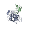
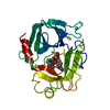
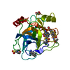
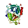
SHEET DETERMINATION METHOD: DSSP THE SHEETS PRESENTED AS "BA" IN EACH CHAIN ON SHEET RECORDS BELOW ... SHEET DETERMINATION METHOD: DSSP THE SHEETS PRESENTED AS "BA" IN EACH CHAIN ON SHEET RECORDS BELOW IS ACTUALLY AN 6-STRANDED BARREL THIS IS REPRESENTED BY A 7-STRANDED SHEET IN WHICH THE FIRST AND LAST STRANDS ARE IDENTICAL.
Mass: 18.015 Da / Num. of mol.: 350 / Source method: isolated from a natural source / Formula: H2O
Sequence details
IN VITRO EVOLVED SEQUENCE FOR CHAIN A.
-
Experimental details
-
Experiment
Experiment
Method: X-RAY DIFFRACTION / Number of used crystals: 1
-
Sample preparation
Crystal
Density Matthews: 1.89 Å3/Da / Density % sol: 34.9 % / Description: NONE
Crystal grow
pH: 4.6 Details: EQUAL AMOUNT OF PROTEIN SOLUTION (9.1 MG/ML PROTEIN COMPLEX IN 0.5 MM MES PH 6.0 BUFFER) AND PRECIPITANT SOLUTION (30% PEG 4000, 0.3 M AMMONIUM ACETATE, 0.1 M NA-ACETATE PH 4.6) WERE MIXED ...Details: EQUAL AMOUNT OF PROTEIN SOLUTION (9.1 MG/ML PROTEIN COMPLEX IN 0.5 MM MES PH 6.0 BUFFER) AND PRECIPITANT SOLUTION (30% PEG 4000, 0.3 M AMMONIUM ACETATE, 0.1 M NA-ACETATE PH 4.6) WERE MIXED AND EQUILIBRATED AGAINST 0.5 ML PRECIPITANT SOLUTION.
Resolution: 0.93→10 Å / Num. parameters: 20959 / Num. restraintsaints: 25983 / Cross valid method: FREE R-VALUE / σ(F): 0 StereochEM target val spec case: ASP-102, HIS-57, SER-195, ASP-194, GLY-193, SER-214 AND SCISSILE PEPTIDE BOND OF INHIBITOR Stereochemistry target values: ENGH AND HUBER Details: ANISOTROPIC REFINEMENT REDUCED FREE R (NO CUTOFF) BY 0.044. THE STRUCTURE WAS REFINED USING UNMERGED REFLECTIONS IN SHELX. THIS DATASET IS INCLUDED WITH THE MAIN STRUCTURE FACTOR FILE ...Details: ANISOTROPIC REFINEMENT REDUCED FREE R (NO CUTOFF) BY 0.044. THE STRUCTURE WAS REFINED USING UNMERGED REFLECTIONS IN SHELX. THIS DATASET IS INCLUDED WITH THE MAIN STRUCTURE FACTOR FILE R2XTTSF AS A SECOND DATASET.
Rfactor
Num. reflection
% reflection
Selection details
Rfree
0.1397
2035
1.6 %
RANDOM
all
0.1168
122128
-
-
obs
-
-
91 %
-
Refine analyze
Num. disordered residues: 21 / Occupancy sum hydrogen: 1820.58 / Occupancy sum non hydrogen: 2225.9
Refinement step
Cycle: LAST / Resolution: 0.93→10 Å
Protein
Nucleic acid
Ligand
Solvent
Total
Num. atoms
1876
0
5
350
2231
Refine LS restraints
Refine-ID
Type
Dev ideal
X-RAY DIFFRACTION
s_bond_d
0.018
X-RAY DIFFRACTION
s_angle_d
0.034
X-RAY DIFFRACTION
s_similar_dist
0
X-RAY DIFFRACTION
s_from_restr_planes
0.0346
X-RAY DIFFRACTION
s_zero_chiral_vol
0.1
X-RAY DIFFRACTION
s_non_zero_chiral_vol
0.107
X-RAY DIFFRACTION
s_anti_bump_dis_restr
0.121
X-RAY DIFFRACTION
s_rigid_bond_adp_cmpnt
0.007
X-RAY DIFFRACTION
s_similar_adp_cmpnt
0.031
X-RAY DIFFRACTION
s_approx_iso_adps
0.116
+
About Yorodumi
-
News
-
Feb 9, 2022. New format data for meta-information of EMDB entries
New format data for meta-information of EMDB entries
Version 3 of the EMDB header file is now the official format.
The previous official version 1.9 will be removed from the archive.
In the structure databanks used in Yorodumi, some data are registered as the other names, "COVID-19 virus" and "2019-nCoV". Here are the details of the virus and the list of structure data.
Jan 31, 2019. EMDB accession codes are about to change! (news from PDBe EMDB page)
EMDB accession codes are about to change! (news from PDBe EMDB page)
The allocation of 4 digits for EMDB accession codes will soon come to an end. Whilst these codes will remain in use, new EMDB accession codes will include an additional digit and will expand incrementally as the available range of codes is exhausted. The current 4-digit format prefixed with “EMD-” (i.e. EMD-XXXX) will advance to a 5-digit format (i.e. EMD-XXXXX), and so on. It is currently estimated that the 4-digit codes will be depleted around Spring 2019, at which point the 5-digit format will come into force.
The EM Navigator/Yorodumi systems omit the EMD- prefix.
Related info.:Q: What is EMD? / ID/Accession-code notation in Yorodumi/EM Navigator
Yorodumi is a browser for structure data from EMDB, PDB, SASBDB, etc.
This page is also the successor to EM Navigator detail page, and also detail information page/front-end page for Omokage search.
The word "yorodu" (or yorozu) is an old Japanese word meaning "ten thousand". "mi" (miru) is to see.
Related info.:EMDB / PDB / SASBDB / Comparison of 3 databanks / Yorodumi Search / Aug 31, 2016. New EM Navigator & Yorodumi / Yorodumi Papers / Jmol/JSmol / Function and homology information / Changes in new EM Navigator and Yorodumi
 Movie
Movie Controller

 Yorodumi
 Open data
 Basic information
 Components
 Keywords
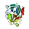
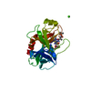
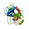
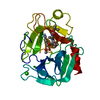
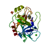
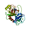
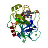
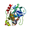
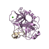
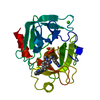
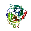
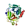
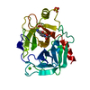
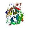
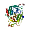
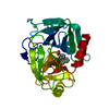
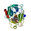
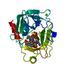
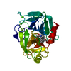
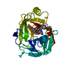
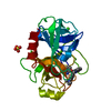
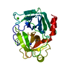
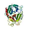
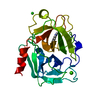
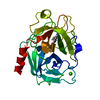
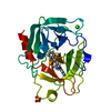
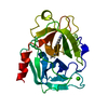
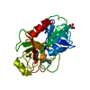
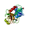
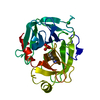
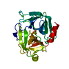
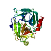
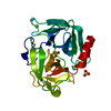
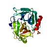
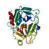
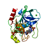
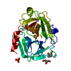
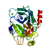
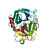
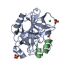
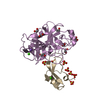
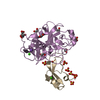
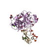
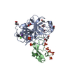
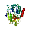
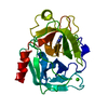
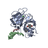
 HYDROLASE /
  Function and homology information

 Authors
 Citation
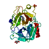
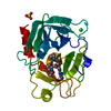
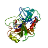
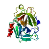
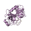
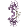
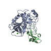
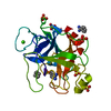
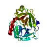
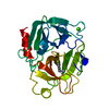
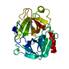
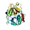
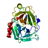
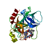
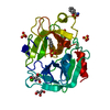
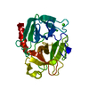
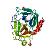
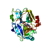
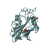
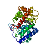
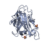
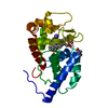
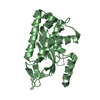
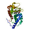
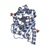
 Structure visualization
 Downloads & links
 Other downloads


 PDBj
PDBj

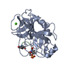
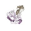
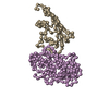
 Assembly


 Mass: 40.078 Da / Num. of mol.: 1 / Source method: obtained synthetically / Formula: Ca
 Mass: 59.044 Da / Num. of mol.: 1 / Source method: obtained synthetically / Formula: C2H3O2
 Mass: 18.015 Da / Num. of mol.: 350 / Source method: isolated from a natural source / Formula: H2O
 Sample preparation
 / Beamline: ID14-1 / Wavelength: 0.9334
  Processing