- PDB-2xqt: Microscopic rotary mechanism of ion translocation in the Fo compl... -
+
Open data
ID or keywords:
Loading...
-
Basic information
Entry
Database: PDB / ID: 2xqt
Title
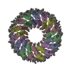
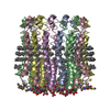
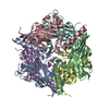
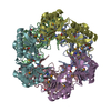
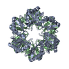
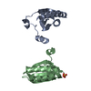
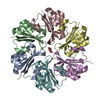
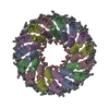
Microscopic rotary mechanism of ion translocation in the Fo complex of ATP synthases
Components
ATP SYNTHASE C CHAIN
Keywords
MEMBRANE PROTEIN / F1FO-ATP SYNTHASE ROTOR / C-RING / ION (PROTON / H+)-TRANSLOCATION / N\ / N'- DICYCLOHEXYLCARBODIIMIDE (DCCD) INHIBITOR
Function / homology
Function and homology information
plasma membrane-derived thylakoid membrane / proton-transporting ATP synthase complex, coupling factor F(o) / proton-transporting ATP synthase activity, rotational mechanism / membrane => GO:0016020 / lipid binding Similarity search - Function
F1F0 ATP synthase subunit C / F1FO ATP Synthase / ATP synthase, F0 complex, subunit C, bacterial/chloroplast / ATP synthase, F0 complex, subunit C / F1F0 ATP synthase subunit C superfamily / ATP synthase, F0 complex, subunit C, DCCD-binding site / ATP synthase c subunit signature. / V-ATPase proteolipid subunit C-like domain / F/V-ATP synthase subunit C superfamily / ATP synthase subunit C ...F1F0 ATP synthase subunit C / F1FO ATP Synthase / ATP synthase, F0 complex, subunit C, bacterial/chloroplast / ATP synthase, F0 complex, subunit C / F1F0 ATP synthase subunit C superfamily / ATP synthase, F0 complex, subunit C, DCCD-binding site / ATP synthase c subunit signature. / V-ATPase proteolipid subunit C-like domain / F/V-ATP synthase subunit C superfamily / ATP synthase subunit C / Up-down Bundle / Mainly Alpha Similarity search - Domain/homology
Group: Atomic model / Derived calculations ...Atomic model / Derived calculations / Non-polymer description / Other / Structure summary / Version format compliance
Mass: 18.015 Da / Num. of mol.: 94 / Source method: isolated from a natural source / Formula: H2O
Nonpolymer details
FME IS NATURAL FORMYL-METHIONINYL N-TERMINAL RESIDUE. GLU 62 IS PARTIALLY MODIFIED BY TREATMENT ...FME IS NATURAL FORMYL-METHIONINYL N-TERMINAL RESIDUE. GLU 62 IS PARTIALLY MODIFIED BY TREATMENT WITH DCCD INHIBITOR TO LEAVE BOUND DCW DICYCLOHEXYLUREA CYMAL-4 DETERGENT (CVM) IS ONLY PARTIALLY RESOLVED OWING TO DISORDER
-
Experimental details
-
Experiment
Experiment
Method: X-RAY DIFFRACTION / Number of used crystals: 1
-
Sample preparation
Crystal
Density Matthews: 3.81 Å3/Da / Density % sol: 67.71 % / Description: NONE
In the structure databanks used in Yorodumi, some data are registered as the other names, "COVID-19 virus" and "2019-nCoV". Here are the details of the virus and the list of structure data.
Jan 31, 2019. EMDB accession codes are about to change! (news from PDBe EMDB page)
EMDB accession codes are about to change! (news from PDBe EMDB page)
The allocation of 4 digits for EMDB accession codes will soon come to an end. Whilst these codes will remain in use, new EMDB accession codes will include an additional digit and will expand incrementally as the available range of codes is exhausted. The current 4-digit format prefixed with “EMD-” (i.e. EMD-XXXX) will advance to a 5-digit format (i.e. EMD-XXXXX), and so on. It is currently estimated that the 4-digit codes will be depleted around Spring 2019, at which point the 5-digit format will come into force.
The EM Navigator/Yorodumi systems omit the EMD- prefix.
Related info.:Q: What is EMD? / ID/Accession-code notation in Yorodumi/EM Navigator
Yorodumi is a browser for structure data from EMDB, PDB, SASBDB, etc.
This page is also the successor to EM Navigator detail page, and also detail information page/front-end page for Omokage search.
The word "yorodu" (or yorozu) is an old Japanese word meaning "ten thousand". "mi" (miru) is to see.
Related info.:EMDB / PDB / SASBDB / Comparison of 3 databanks / Yorodumi Search / Aug 31, 2016. New EM Navigator & Yorodumi / Yorodumi Papers / Jmol/JSmol / Function and homology information / Changes in new EM Navigator and Yorodumi
 Movie
Movie Controller

 Yorodumi
 Open data
 Basic information
 Components
 Keywords
 MEMBRANE PROTEIN / F1FO-ATP SYNTHASE ROTOR /
  Function and homology information
 Authors
 Citation
 Structure visualization
 Downloads & links
 Other downloads
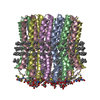


 PDBj
PDBj

 Assembly
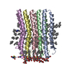

 Mass: 224.342 Da / Num. of mol.: 5 / Source method: obtained synthetically / Formula: C13H24N2O
 Mass: 480.546 Da / Num. of mol.: 45 / Source method: obtained synthetically / Formula: C22H40O11
 Mass: 18.015 Da / Num. of mol.: 94 / Source method: isolated from a natural source / Formula: H2O
 Sample preparation
 / Beamline: X10SA / Wavelength: 0.9999
  Processing