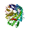
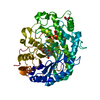
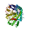
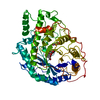
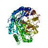
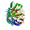
DETERMINATION METHOD: DSSP THE SHEETS PRESENTED AS "AA" IN EACH CHAIN ON SHEET RECORDS BELOW IS ... DETERMINATION METHOD: DSSP THE SHEETS PRESENTED AS "AA" IN EACH CHAIN ON SHEET RECORDS BELOW IS ACTUALLY AN 8-STRANDED BARREL THIS IS REPRESENTED BY A 9-STRANDED SHEET IN WHICH THE FIRST AND LAST STRANDS ARE IDENTICAL.
Mass: 18.015 Da / Num. of mol.: 948 / Source method: isolated from a natural source / Formula: H2O
Nonpolymer details
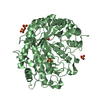
1,2-ETHANEDIOL (EDO): PRESENT AT 20 PERCENT IN THE CRYOPROTECTANT
Sequence details
PROTEIN ISOLATED FROM NATURAL SOURCE BUT SEQUENCE DIFFERS AT 3 POSITIONS TO UNIPROTKB DATABASE ...PROTEIN ISOLATED FROM NATURAL SOURCE BUT SEQUENCE DIFFERS AT 3 POSITIONS TO UNIPROTKB DATABASE ENTRY P16098. THESE CHANGES WERE IDENTIFIED FROM ELECTRON DENSITY MAPS.
-
Experimental details
-
Experiment
Experiment
Method: X-RAY DIFFRACTION / Number of used crystals: 1
-
Sample preparation
Crystal
Density Matthews: 1.91 Å3/Da / Density % sol: 35.6 % / Description: NONE
Crystal grow
Temperature: 291 K / Method: vapor diffusion, hanging drop / pH: 5.5 Details: CRYSTALS WERE GROWN AT 291 K USING THE HANGING DROP VAPOUR DIFFUSION METHOD WITH PROTEIN AT 10 MG PER ML AND A PRECIPITANT COMPRISED OF 14 PERCENT PEG 3350 IN 100 MM BIS-TRIS PROPANE BUFFER AT PH 5.5
 Movie
Movie Controller


 Open data
 Basic information
 Components
 Keywords
 Function and homology information
 Authors
 Citation
 Structure visualization
 Downloads & links
 Other downloads


 PDBj
PDBj Assembly
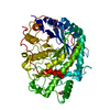

 Mass: 62.068 Da / Num. of mol.: 2 / Source method: obtained synthetically / Formula: C2H6O2
 Mass: 18.015 Da / Num. of mol.: 948 / Source method: isolated from a natural source / Formula: H2O
 Sample preparation
 / Beamline: I02 / Wavelength: 0.9507
  Processing