 Movie
Movie Controller

[English] 日本語
 Yorodumi
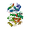
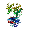
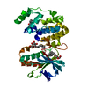
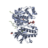
- PDB-2x7g: Structure of human serine-arginine-rich protein-specific kinase 2... -
+ Open data

- Basic information
| Entry | Database: PDB / ID: 2x7g | ||||||
|---|---|---|---|---|---|---|---|
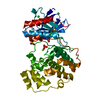
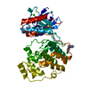
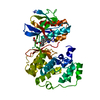
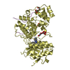
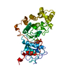
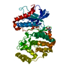
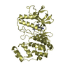
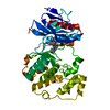
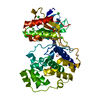
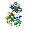
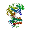
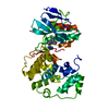
| Title | Structure of human serine-arginine-rich protein-specific kinase 2 (SRPK2) bound to purvalanol B | ||||||
 Components Components | UNCHARACTERIZED PROTEIN SRPK2 | ||||||
 Keywords Keywords |  TRANSFERASE / SERINE/THREONINE-PROTEIN KINASE / MRNA SPLICING / PHOSPHOPROTEIN / MRNA PROCESSING / DIFFERENTIATION / NUCLEOTIDE-BINDING / SPLICEASOME ASSEMBLY / ATP COMPETITIVE INHIBITOR / KINASE / NUCLEUS / ATP-BINDING / SPLICE FACTOR TRAFFICKING TRANSFERASE / SERINE/THREONINE-PROTEIN KINASE / MRNA SPLICING / PHOSPHOPROTEIN / MRNA PROCESSING / DIFFERENTIATION / NUCLEOTIDE-BINDING / SPLICEASOME ASSEMBLY / ATP COMPETITIVE INHIBITOR / KINASE / NUCLEUS / ATP-BINDING / SPLICE FACTOR TRAFFICKING | ||||||
| Function / homology |  Function and homology information Function and homology informationnuclear speck organization / R-loop processing / regulation of mRNA processing / regulation of mRNA splicing, via spliceosome / Maturation of nucleoprotein / spliceosomal complex assembly / negative regulation of viral genome replication / positive regulation of viral genome replication / positive regulation of cell cycle / RNA splicing ...nuclear speck organization / R-loop processing / regulation of mRNA processing / regulation of mRNA splicing, via spliceosome / Maturation of nucleoprotein / spliceosomal complex assembly / negative regulation of viral genome replication / positive regulation of viral genome replication / positive regulation of cell cycle / RNA splicing / 14-3-3 protein binding / positive regulation of neuron apoptotic process / angiogenesis / peptidyl-serine phosphorylation / cell differentiation / non-specific serine/threonine protein kinase / intracellular signal transduction / nuclear speck / protein phosphorylation / innate immune response / protein serine kinase activity / protein serine/threonine kinase activity / positive regulation of cell population proliferation / chromatin / positive regulation of gene expression / nucleolus / magnesium ion binding / RNA binding / nucleoplasm / ATP binding / nucleus / cytosol / cytoplasmSimilarity search - Function | ||||||
| Biological species |  HOMO SAPIENS (human) HOMO SAPIENS (human) | ||||||
| Method | X-RAY DIFFRACTION / SYNCHROTRON / MOLECULAR REPLACEMENT / Resolution: 2.5 Å | ||||||
 Authors Authors | Pike, A.C.W. / Savitsky, P. / Fedorov, O. / Krojer, T. / Ugochukwu, E. / von Delft, F. / Gileadi, O. / Edwards, A. / Arrowsmith, C.H. / Weigelt, J. ...Pike, A.C.W. / Savitsky, P. / Fedorov, O. / Krojer, T. / Ugochukwu, E. / von Delft, F. / Gileadi, O. / Edwards, A. / Arrowsmith, C.H. / Weigelt, J. / Bountra, C. / Knapp, S. | ||||||
 Citation Citation | Journal: To be Published Title: Structure of Human Serine-Arginine-Rich Protein- Specific Kinase 2 (Srpk2) Bound to Purvalanol B Authors: Pike, A.C.W. / Savitsky, P. / Fedorov, O. / Krojer, T. / Ugochukwu, E. / von Delft, F. / Gileadi, O. / Edwards, A. / Arrowsmith, C.H. / Weigelt, J. / Bountra, C. / Knapp, S. | ||||||
| History |
|
- Structure visualization
| Structure viewer | Molecule: MolmilJmol/JSmol |
|---|
- Downloads & links
-Download
| PDBx/mmCIF format | 2x7g.cif.gz | 90.3 KB | Display | PDBx/mmCIF format |
|---|---|---|---|---|
| PDB format | pdb2x7g.ent.gz | 67 KB | Display | PDB format |
| PDBx/mmJSON format | 2x7g.json.gz | Tree view | PDBx/mmJSON format | |
| Others |  Other downloads Other downloads |
-Validation report
| Arichive directory | https://data.pdbj.org/pub/pdb/validation_reports/x7/2x7gftp://data.pdbj.org/pub/pdb/validation_reports/x7/2x7g | HTTPS FTP |
|---|
-Related structure data
| Related structure data |  1wbpS S: Starting model for refinement |
|---|---|
| Similar structure data |


-Links
 PDBj
PDBj- Assembly
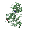
| Deposited unit | 
| ||||||||
|---|---|---|---|---|---|---|---|---|---|
| 1 |
| ||||||||
| Unit cell |
|
-Components
-Protein , 1 types, 1 molecules A
| #1: Protein | Mass: 44300.676 Da / Num. of mol.: 1 / Fragment: KINASE DOMAIN, RESIDUES 62-267,519-699 Source method: isolated from a genetically manipulated source Details: SYNTHETIC CONSTRUCT ENGINEERED TO REMOVE INTERVENING REGION. Source: (gene. exp.) HOMO SAPIENS (human) / Plasmid: PNIC28-BSA4 / Production host:  ESCHERICHIA COLI (E. coli) / Strain (production host): BL21(DE3) / Variant (production host): R3-PRARE2 ESCHERICHIA COLI (E. coli) / Strain (production host): BL21(DE3) / Variant (production host): R3-PRARE2References: UniProt: C9JQJ0, UniProt: P78362*PLUS, non-specific serine/threonine protein kinase |
|---|
-Non-polymers , 5 types, 126 molecules 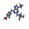


| #2: Chemical | ChemComp-PVB / Cyclin-dependent kinase 2 Mass: 432.904 Da / Num. of mol.: 1 / Source method: obtained synthetically / Formula: C20H25ClN6O3 Mass: 432.904 Da / Num. of mol.: 1 / Source method: obtained synthetically / Formula: C20H25ClN6O3 | ||||||
|---|---|---|---|---|---|---|---|
| #3: Chemical | ChemComp-SO4 / Sulfate Mass: 96.063 Da / Num. of mol.: 7 / Source method: obtained synthetically / Formula: SO4 Mass: 96.063 Da / Num. of mol.: 7 / Source method: obtained synthetically / Formula: SO4#4: Chemical | ChemComp-ACT / | Acetate Mass: 59.044 Da / Num. of mol.: 1 / Source method: obtained synthetically / Formula: C2H3O2 Mass: 59.044 Da / Num. of mol.: 1 / Source method: obtained synthetically / Formula: C2H3O2#5: Chemical | ChemComp-EDO / Ethylene glycol Mass: 62.068 Da / Num. of mol.: 6 / Source method: obtained synthetically / Formula: C2H6O2 Mass: 62.068 Da / Num. of mol.: 6 / Source method: obtained synthetically / Formula: C2H6O2#6: Water | ChemComp-HOH / | WaterMass: 18.015 Da / Num. of mol.: 111 / Source method: isolated from a natural source / Formula: H2O |
-Details
| Sequence details | SYNTHETIC CONSTRUCT WITH 268-518 REMOVED |
|---|
-Experimental details
-Experiment
| Experiment | Method: X-RAY DIFFRACTION / Number of used crystals: 1 |
|---|
- Sample preparation
| Crystal | Density Matthews: 3.6 Å3/Da / Density % sol: 65.82 % / Description: NONE |
|---|---|
| Crystal grow | pH: 4.75 / Details: 0.5M AMMONIUM SULPHATE, 0.1M SODIUM ACETATE PH4.75 |
-Data collection
| Diffraction | Mean temperature: 100 K |
|---|---|
| Diffraction source | Source: SYNCHROTRON / Site: Diamond  / Beamline: I24 / Wavelength: 0.9796 / Beamline: I24 / Wavelength: 0.9796 |
| Detector | Type: RAYONIX / Detector: CCD / Date: Sep 28, 2009 |
| Radiation | Protocol: SINGLE WAVELENGTH / Monochromatic (M) / Laue (L): M / Scattering type: x-ray |
| Radiation wavelength | Wavelength: 0.9796 Å / Relative weight: 1 |
| Reflection | Resolution: 2.5→45.21 Å / Num. obs: 22306 / % possible obs: 99.7 % / Observed criterion σ(I): 0 / Redundancy: 3.2 % / Biso Wilson estimate: 43.1 Å2 / Rmerge(I) obs: 0.13 / Net I/σ(I): 6.5 |
| Reflection shell | Resolution: 2.5→2.64 Å / Redundancy: 3.2 % / Rmerge(I) obs: 0.59 / Mean I/σ(I) obs: 2 / % possible all: 99.6 |
- Processing
| Software |
| ||||||||||||||||||||||||||||||||||||||||||||||||||||||||||||||||||||||||||||||||||||||||||||||||||||||||||||||||||||||||||||||||||||||||||||||||||||||||||||||||||||||||||||||||||||||
|---|---|---|---|---|---|---|---|---|---|---|---|---|---|---|---|---|---|---|---|---|---|---|---|---|---|---|---|---|---|---|---|---|---|---|---|---|---|---|---|---|---|---|---|---|---|---|---|---|---|---|---|---|---|---|---|---|---|---|---|---|---|---|---|---|---|---|---|---|---|---|---|---|---|---|---|---|---|---|---|---|---|---|---|---|---|---|---|---|---|---|---|---|---|---|---|---|---|---|---|---|---|---|---|---|---|---|---|---|---|---|---|---|---|---|---|---|---|---|---|---|---|---|---|---|---|---|---|---|---|---|---|---|---|---|---|---|---|---|---|---|---|---|---|---|---|---|---|---|---|---|---|---|---|---|---|---|---|---|---|---|---|---|---|---|---|---|---|---|---|---|---|---|---|---|---|---|---|---|---|---|---|---|---|
| Refinement | Method to determine structure: MOLECULAR REPLACEMENT Starting model: PDB ENTRY 1WBP Resolution: 2.5→45 Å / Cor.coef. Fo:Fc: 0.943 / Cor.coef. Fo:Fc free: 0.893 / SU B: 12.616 / SU ML: 0.148 / TLS residual ADP flag: LIKELY RESIDUAL / Cross valid method: THROUGHOUT / ESU R: 0.263 / ESU R Free: 0.237 / Stereochemistry target values: MAXIMUM LIKELIHOOD Details: HYDROGENS HAVE BEEN ADDED IN THE RIDING POSITIONS. ATOM RECORD CONTAINS RESIDUAL B FACTORS ONLY
| ||||||||||||||||||||||||||||||||||||||||||||||||||||||||||||||||||||||||||||||||||||||||||||||||||||||||||||||||||||||||||||||||||||||||||||||||||||||||||||||||||||||||||||||||||||||
| Solvent computation | Ion probe radii: 0.8 Å / Shrinkage radii: 0.8 Å / VDW probe radii: 1.4 Å / Solvent model: MASK | ||||||||||||||||||||||||||||||||||||||||||||||||||||||||||||||||||||||||||||||||||||||||||||||||||||||||||||||||||||||||||||||||||||||||||||||||||||||||||||||||||||||||||||||||||||||
| Displacement parameters | Biso mean: 16.352 Å2
| ||||||||||||||||||||||||||||||||||||||||||||||||||||||||||||||||||||||||||||||||||||||||||||||||||||||||||||||||||||||||||||||||||||||||||||||||||||||||||||||||||||||||||||||||||||||
| Refinement step | Cycle: LAST / Resolution: 2.5→45 Å
| ||||||||||||||||||||||||||||||||||||||||||||||||||||||||||||||||||||||||||||||||||||||||||||||||||||||||||||||||||||||||||||||||||||||||||||||||||||||||||||||||||||||||||||||||||||||
| Refine LS restraints |
|