 Movie
Movie Controller


+ Open data

- Basic information
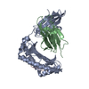
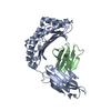
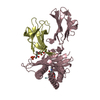
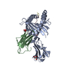
| Entry | Database: PDB / ID: 2x79 | ||||||
|---|---|---|---|---|---|---|---|
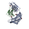
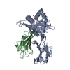
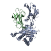
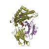
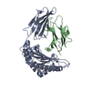
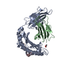
| Title | Inward facing conformation of Mhp1 | ||||||
 Components Components | HYDANTOIN TRANSPORT PROTEIN | ||||||
 Keywords Keywords |  TRANSPORT PROTEIN / TRANSPORTER / MEMBRANE PROTEIN TRANSPORT PROTEIN / TRANSPORTER / MEMBRANE PROTEIN | ||||||
| Function / homology |  Function and homology information Function and homology information | ||||||
| Biological species |  MICROBACTERIUM LIQUEFACIENS (bacteria) MICROBACTERIUM LIQUEFACIENS (bacteria) | ||||||
| Method | X-RAY DIFFRACTION / SYNCHROTRON / SAD / Resolution: 3.8 Å | ||||||
 Authors Authors | Shimamura, T. / Weyand, S. / Beckstein, O. / Rutherford, N.G. / Hadden, J.M. / Sharples, D. / Sansom, M.S.P. / Iwata, S. / Henderson, P.J.F. / Cameron, A.D. | ||||||
 Citation Citation | Journal: Science / Year: 2010 Title: Molecular Basis of Alternating Access Membrane Transport by the Sodium-Hydantoin Transporter Mhp1. Authors: Shimamura, T. / Weyand, S. / Beckstein, O. / Rutherford, N.G. / Hadden, J.M. / Sharples, D. / Sansom, M.S.P. / Iwata, S. / Henderson, P.J.F. / Cameron, A.D. | ||||||
| History |
|
- Structure visualization
| Structure viewer | Molecule: MolmilJmol/JSmol |
|---|
- Downloads & links
-Download
| PDBx/mmCIF format | 2x79.cif.gz | 191.3 KB | Display | PDBx/mmCIF format |
|---|---|---|---|---|
| PDB format | pdb2x79.ent.gz | 165 KB | Display | PDB format |
| PDBx/mmJSON format | 2x79.json.gz | Tree view | PDBx/mmJSON format | |
| Others |  Other downloads Other downloads |
-Validation report
| Arichive directory | https://data.pdbj.org/pub/pdb/validation_reports/x7/2x79ftp://data.pdbj.org/pub/pdb/validation_reports/x7/2x79 | HTTPS FTP |
|---|
-Related structure data
| Similar structure data |
|---|

-Links
 PDBj
PDBj- Assembly
| Deposited unit | 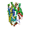
| ||||||||
|---|---|---|---|---|---|---|---|---|---|
| 1 |
| ||||||||
| Unit cell |
|
-Components
| #1: Protein | Mass: 55556.680 Da / Num. of mol.: 1 Source method: isolated from a genetically manipulated source Details: SELENOMETHIONINE / Source: (gene. exp.) MICROBACTERIUM LIQUEFACIENS (bacteria) / Strain: AJ3912 / Plasmid: PSHP11 / Production host: ESCHERICHIA COLI (E. coli) / Strain (production host): BLR / References: UniProt: D6R8X8*PLUS |
|---|---|
| Sequence details | NOT PUBLISHED |
-Experimental details
-Experiment
| Experiment | Method: X-RAY DIFFRACTION / Number of used crystals: 1 |
|---|
- Sample preparation
| Crystal | Density Matthews: 7.2 Å3/Da / Density % sol: 83 % / Description: BUILDING WAS BASED ON PDB ENTRY 2JLN |
|---|---|
| Crystal grow | pH: 9 / Details: 24-28% PEG300, 100 MM NACL AND 100 MM BICINE PH9.0 |
-Data collection
| Diffraction | Mean temperature: 100 K |
|---|---|
| Diffraction source | Source: SYNCHROTRON / Site: ESRF  / Beamline: ID29 / Wavelength: 0.979147 / Beamline: ID29 / Wavelength: 0.979147 |
| Detector | Type: ADSC QUANTUM 315 / Detector: CCD / Date: Jun 13, 2004 / Details: MIRRORS |
| Radiation | Monochromator: SI(111) / Protocol: SINGLE WAVELENGTH / Monochromatic (M) / Laue (L): M / Scattering type: x-ray |
| Radiation wavelength | Wavelength: 0.979147 Å / Relative weight: 1 |
| Reflection | Resolution: 3.8→40 Å / Num. obs: 47973 / % possible obs: 97 % / Observed criterion σ(I): -1 / Redundancy: 3.8 % / Biso Wilson estimate: 172.82 Å2 / Rmerge(I) obs: 0.08 / Net I/σ(I): 17.6 |
| Reflection shell | Resolution: 3.8→3.94 Å / Redundancy: 2.8 % / Rmerge(I) obs: 0.69 / Mean I/σ(I) obs: 1.4 / % possible all: 96 |
- Processing
| Software |
| |||||||||||||||||||||||||||||||||||||||||||||||||||||||||||||||||||||||||||||||||||||||||||||||||||||||||||||||||||||||||||||
|---|---|---|---|---|---|---|---|---|---|---|---|---|---|---|---|---|---|---|---|---|---|---|---|---|---|---|---|---|---|---|---|---|---|---|---|---|---|---|---|---|---|---|---|---|---|---|---|---|---|---|---|---|---|---|---|---|---|---|---|---|---|---|---|---|---|---|---|---|---|---|---|---|---|---|---|---|---|---|---|---|---|---|---|---|---|---|---|---|---|---|---|---|---|---|---|---|---|---|---|---|---|---|---|---|---|---|---|---|---|---|---|---|---|---|---|---|---|---|---|---|---|---|---|---|---|---|
| Refinement | Method to determine structure: SAD Starting model: NONE Resolution: 3.8→33.388 Å / SU ML: 0.83 / σ(F): 1.96 / Phase error: 39.94 / Stereochemistry target values: ML Details: MODELLING WAS DONE WITH SHARPENED MAPS. IN THE SODIUM BINDING SITE BETWEEN RESIDUES ALA38 AND THR330 THERE IS ADDITIONAL DENSITY THAT HAS NOT BEEN MODELLED.
| |||||||||||||||||||||||||||||||||||||||||||||||||||||||||||||||||||||||||||||||||||||||||||||||||||||||||||||||||||||||||||||
| Solvent computation | Shrinkage radii: 0.9 Å / VDW probe radii: 1.11 Å / Solvent model: FLAT BULK SOLVENT MODEL / Bsol: 99 Å2 / ksol: 0.262 e/Å3 | |||||||||||||||||||||||||||||||||||||||||||||||||||||||||||||||||||||||||||||||||||||||||||||||||||||||||||||||||||||||||||||
| Displacement parameters | Biso mean: 242 Å2
| |||||||||||||||||||||||||||||||||||||||||||||||||||||||||||||||||||||||||||||||||||||||||||||||||||||||||||||||||||||||||||||
| Refinement step | Cycle: LAST / Resolution: 3.8→33.388 Å
| |||||||||||||||||||||||||||||||||||||||||||||||||||||||||||||||||||||||||||||||||||||||||||||||||||||||||||||||||||||||||||||
| Refine LS restraints |
| |||||||||||||||||||||||||||||||||||||||||||||||||||||||||||||||||||||||||||||||||||||||||||||||||||||||||||||||||||||||||||||
| LS refinement shell |
| |||||||||||||||||||||||||||||||||||||||||||||||||||||||||||||||||||||||||||||||||||||||||||||||||||||||||||||||||||||||||||||
| Refinement TLS params. | Method: refined / Refine-ID: X-RAY DIFFRACTION
| |||||||||||||||||||||||||||||||||||||||||||||||||||||||||||||||||||||||||||||||||||||||||||||||||||||||||||||||||||||||||||||
| Refinement TLS group |
|
