 Movie
Movie Controller

[English] 日本語
 Yorodumi
- PDB-2wxg: The crystal structure of the murine class IA PI 3-kinase p110delt... -
+ Open data

- Basic information
| Entry | Database: PDB / ID: 2wxg | ||||||
|---|---|---|---|---|---|---|---|
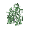
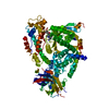
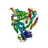
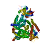
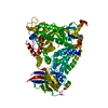
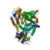
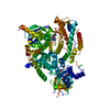
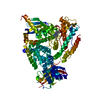
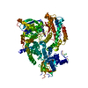
| Title | The crystal structure of the murine class IA PI 3-kinase p110delta in complex with SW13. | ||||||
 Components Components | PHOSPHATIDYLINOSITOL-4,5-BISPHOSPHATE 3-KINASE CATALYTIC SUBUNIT DELTA ISOFORM | ||||||
 Keywords Keywords |  TRANSFERASE / PHOSPHOPROTEIN / ISOFORM-SPECIFIC INHIBITORS / CANCER TRANSFERASE / PHOSPHOPROTEIN / ISOFORM-SPECIFIC INHIBITORS / CANCER | ||||||
| Function / homology |  Function and homology information Function and homology informationErythropoietin activates Phosphoinositide-3-kinase (PI3K) / Interleukin receptor SHC signaling / Synthesis of PIPs at the plasma membrane / PIP3 activates AKT signaling / Antigen activates B Cell Receptor (BCR) leading to generation of second messengers / Regulation of signaling by CBL / positive regulation of cell migration by vascular endothelial growth factor signaling pathway / PI5P, PP2A and IER3 Regulate PI3K/AKT Signaling / Interleukin-3, Interleukin-5 and GM-CSF signaling / RET signaling ...Erythropoietin activates Phosphoinositide-3-kinase (PI3K) / Interleukin receptor SHC signaling / Synthesis of PIPs at the plasma membrane / PIP3 activates AKT signaling / Antigen activates B Cell Receptor (BCR) leading to generation of second messengers / Regulation of signaling by CBL / positive regulation of cell migration by vascular endothelial growth factor signaling pathway / PI5P, PP2A and IER3 Regulate PI3K/AKT Signaling / Interleukin-3, Interleukin-5 and GM-CSF signaling / RET signaling / positive regulation of epithelial tube formation / positive regulation of neutrophil apoptotic process / phosphatidylinositol-mediated signaling / phosphatidylinositol 3-kinase complex / 1-phosphatidylinositol-4-phosphate 3-kinase activity / 1-phosphatidylinositol-4,5-bisphosphate 3-kinase activity / phosphatidylinositol-4,5-bisphosphate 3-kinase / phosphatidylinositol 3-kinase / phosphatidylinositol 3-kinase complex, class IA / phosphatidylinositol-3-phosphate biosynthetic process / 1-phosphatidylinositol-3-kinase activity / B cell activation / B cell homeostasis / homeostasis of number of cells / defense response to fungus / positive regulation of endothelial cell proliferation / positive regulation of endothelial cell migration / phosphatidylinositol 3-kinase/protein kinase B signal transduction / positive regulation of angiogenesis / chemotaxis / kinase activity / adaptive immune response / cell differentiation / cell surface receptor signaling pathway / positive regulation of cell migration / inflammatory response / phosphorylation / negative regulation of gene expression / innate immune response / positive regulation of gene expression / ATP binding / plasma membrane / cytosol / cytoplasmSimilarity search - Function | ||||||
| Biological species |  MUS MUSCULUS (house mouse) MUS MUSCULUS (house mouse) | ||||||
| Method | X-RAY DIFFRACTION / SYNCHROTRON / MOLECULAR REPLACEMENT / Resolution: 2 Å | ||||||
 Authors Authors | Berndt, A. / Miller, S. / Williams, O. / Lee, D.D. / Houseman, B.T. / Pacold, J.I. / Gorrec, F. / Hon, W.-C. / Liu, Y. / Rommel, C. ...Berndt, A. / Miller, S. / Williams, O. / Lee, D.D. / Houseman, B.T. / Pacold, J.I. / Gorrec, F. / Hon, W.-C. / Liu, Y. / Rommel, C. / Gaillard, P. / Ruckle, T. / Schwarz, M.K. / Shokat, K.M. / Shaw, J.P. / Williams, R.L. | ||||||
 Citation Citation | Journal: Nat.Chem.Biol. / Year: 2010 Title: The P110D Structure: Mechanisms for Selectivity and Potency of New Pi(3)K Inhibitors Authors: Berndt, A. / Miller, S. / Williams, O. / Lee, D.D. / Houseman, B.T. / Pacold, J.I. / Gorrec, F. / Hon, W.-C. / Liu, Y. / Rommel, C. / Gaillard, P. / Ruckle, T. / Schwarz, M.K. / Shokat, K.M. ...Authors: Berndt, A. / Miller, S. / Williams, O. / Lee, D.D. / Houseman, B.T. / Pacold, J.I. / Gorrec, F. / Hon, W.-C. / Liu, Y. / Rommel, C. / Gaillard, P. / Ruckle, T. / Schwarz, M.K. / Shokat, K.M. / Shaw, J.P. / Williams, R.L. | ||||||
| History |
| ||||||
| Remark 700 | SHEET THE SHEET STRUCTURE OF THIS MOLECULE IS BIFURCATED. IN ORDER TO REPRESENT THIS FEATURE IN ... SHEET THE SHEET STRUCTURE OF THIS MOLECULE IS BIFURCATED. IN ORDER TO REPRESENT THIS FEATURE IN THE SHEET RECORDS BELOW, TWO SHEETS ARE DEFINED. |
- Structure visualization
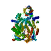
| Structure viewer | Molecule: MolmilJmol/JSmol |
|---|
- Downloads & links
-Download
| PDBx/mmCIF format | 2wxg.cif.gz | 187.9 KB | Display | PDBx/mmCIF format |
|---|---|---|---|---|
| PDB format | pdb2wxg.ent.gz | 143.7 KB | Display | PDB format |
| PDBx/mmJSON format | 2wxg.json.gz | Tree view | PDBx/mmJSON format | |
| Others |  Other downloads Other downloads |
-Validation report
| Arichive directory | https://data.pdbj.org/pub/pdb/validation_reports/wx/2wxgftp://data.pdbj.org/pub/pdb/validation_reports/wx/2wxg | HTTPS FTP |
|---|
-Related structure data
| Related structure data |  2wxfC  2wxhC  2wxiC  2wxjC  2wxkC  2wxlC  2wxmC  2wxnC  2wxoC  2wxpC  2wxqC  2wxrC  2x38C  2rd0S C: citing same article ( S: Starting model for refinement |
|---|---|
| Similar structure data |


-Links
 PDBj
PDBj


- Assembly
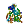
| Deposited unit | 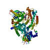
| ||||||||
|---|---|---|---|---|---|---|---|---|---|
| 1 |
| ||||||||
| Unit cell |
|
-Components
| #1: Protein | Mass: 107823.664 Da / Num. of mol.: 1 / Fragment: RESIDUES 106-1044 Source method: isolated from a genetically manipulated source Source: (gene. exp.) MUS MUSCULUS (house mouse) / Plasmid: PFASTBAC HTA / Cell line (production host): Sf21 / Production host:   SPODOPTERA FRUGIPERDA (fall armyworm) SPODOPTERA FRUGIPERDA (fall armyworm)References: UniProt: Q3UDT3, UniProt: O35904*PLUS, phosphatidylinositol-4,5-bisphosphate 3-kinase |
|---|---|
| #2: Chemical | ChemComp-ZZN /   Mass: 507.518 Da / Num. of mol.: 1 / Source method: obtained synthetically / Formula: C28H22FN7O2 Mass: 507.518 Da / Num. of mol.: 1 / Source method: obtained synthetically / Formula: C28H22FN7O2 |
| #3: Water | ChemComp-HOH / Water Mass: 18.015 Da / Num. of mol.: 187 / Source method: isolated from a natural source / Formula: H2O Mass: 18.015 Da / Num. of mol.: 187 / Source method: isolated from a natural source / Formula: H2O |
-Experimental details
-Experiment
| Experiment | Method: X-RAY DIFFRACTION / Number of used crystals: 1 |
|---|
- Sample preparation
| Crystal | Density Matthews: 2.44 Å3/Da / Density % sol: 49.55 % / Description: NONE |
|---|---|
| Crystal grow | pH: 6.8 Details: 20% (V/V) GLYCEROL, 10% (W/V) PEG 4K, 30 MM NANO3, 30 MM NA2HPO4, 30 MM (NH4)2SO4, 100 MM IMIDAZOLE PH 6.8 |
-Data collection
| Diffraction | Mean temperature: 100 K |
|---|---|
| Diffraction source | Source: SYNCHROTRON / Site: ESRF  / Beamline: ID14-4 / Wavelength: 0.975 / Beamline: ID14-4 / Wavelength: 0.975 |
| Detector | Type: ADSC CCD / Detector: CCD / Date: Jun 28, 2008 |
| Radiation | Protocol: SINGLE WAVELENGTH / Monochromatic (M) / Laue (L): M / Scattering type: x-ray |
| Radiation wavelength | Wavelength: 0.975 Å / Relative weight: 1 |
| Reflection | Resolution: 2→69.01 Å / Num. obs: 69666 / % possible obs: 99.8 % / Observed criterion σ(I): -3.7 / Redundancy: 7.38 % / Biso Wilson estimate: 31.443 Å2 / Rmerge(I) obs: 0.09 / Net I/σ(I): 15.69 |
| Reflection shell | Resolution: 2→2.02 Å / Redundancy: 7.44 % / Rmerge(I) obs: 0.72 / Mean I/σ(I) obs: 1.82 / % possible all: 100 |
- Processing
| Software |
| ||||||||||||||||||||||||||||||||||||||||||||||||||||||||||||||||||||||||||||||||||||||||||||||||||||||||||||||||||||||||||||||||||||||||||||||||||||||||||||||||||||||||||||||||||||||
|---|---|---|---|---|---|---|---|---|---|---|---|---|---|---|---|---|---|---|---|---|---|---|---|---|---|---|---|---|---|---|---|---|---|---|---|---|---|---|---|---|---|---|---|---|---|---|---|---|---|---|---|---|---|---|---|---|---|---|---|---|---|---|---|---|---|---|---|---|---|---|---|---|---|---|---|---|---|---|---|---|---|---|---|---|---|---|---|---|---|---|---|---|---|---|---|---|---|---|---|---|---|---|---|---|---|---|---|---|---|---|---|---|---|---|---|---|---|---|---|---|---|---|---|---|---|---|---|---|---|---|---|---|---|---|---|---|---|---|---|---|---|---|---|---|---|---|---|---|---|---|---|---|---|---|---|---|---|---|---|---|---|---|---|---|---|---|---|---|---|---|---|---|---|---|---|---|---|---|---|---|---|---|---|
| Refinement | Method to determine structure: MOLECULAR REPLACEMENT Starting model: PDB ENTRY 2RD0 Resolution: 2→113.96 Å / Cor.coef. Fo:Fc: 0.944 / Cor.coef. Fo:Fc free: 0.922 / SU B: 8.259 / SU ML: 0.109 / TLS residual ADP flag: LIKELY RESIDUAL / Cross valid method: THROUGHOUT / ESU R: 0.175 / ESU R Free: 0.158 / Stereochemistry target values: MAXIMUM LIKELIHOOD / Details: HYDROGENS HAVE BEEN ADDED IN THE RIDING POSITIONS.
| ||||||||||||||||||||||||||||||||||||||||||||||||||||||||||||||||||||||||||||||||||||||||||||||||||||||||||||||||||||||||||||||||||||||||||||||||||||||||||||||||||||||||||||||||||||||
| Solvent computation | Ion probe radii: 0.8 Å / Shrinkage radii: 0.8 Å / VDW probe radii: 1.2 Å / Solvent model: MASK | ||||||||||||||||||||||||||||||||||||||||||||||||||||||||||||||||||||||||||||||||||||||||||||||||||||||||||||||||||||||||||||||||||||||||||||||||||||||||||||||||||||||||||||||||||||||
| Displacement parameters | Biso mean: 24.549 Å2
| ||||||||||||||||||||||||||||||||||||||||||||||||||||||||||||||||||||||||||||||||||||||||||||||||||||||||||||||||||||||||||||||||||||||||||||||||||||||||||||||||||||||||||||||||||||||
| Refinement step | Cycle: LAST / Resolution: 2→113.96 Å
| ||||||||||||||||||||||||||||||||||||||||||||||||||||||||||||||||||||||||||||||||||||||||||||||||||||||||||||||||||||||||||||||||||||||||||||||||||||||||||||||||||||||||||||||||||||||
| Refine LS restraints |
|