Mass: 18.015 Da / Num. of mol.: 198 / Source method: isolated from a natural source / Formula: H2O
Compound details
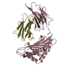
ENGINEERED RESIDUE IN CHAIN A, ASP 23 TO ALA ENGINEERED RESIDUE IN CHAIN A, PRO 33 TO HIS ...ENGINEERED RESIDUE IN CHAIN A, ASP 23 TO ALA ENGINEERED RESIDUE IN CHAIN A, PRO 33 TO HIS ENGINEERED RESIDUE IN CHAIN B, ASP 23 TO ALA ENGINEERED RESIDUE IN CHAIN B, PRO 33 TO HIS
-
Experimental details
-
Experiment
Experiment
Method: X-RAY DIFFRACTION / Number of used crystals: 1
-
Sample preparation
Crystal
Density Matthews: 2.67 Å3/Da / Density % sol: 53.57 % / Description: NONE
Crystal grow
Temperature: 276 K / Details: CRYSTALLIZED AT 276K IN 25% ETHYLENE GLYCOL
Resolution: 2→65.23 Å / Cor.coef. Fo:Fc: 0.944 / Cor.coef. Fo:Fc free: 0.917 / SU B: 12.51 / SU ML: 0.182 / TLS residual ADP flag: LIKELY RESIDUAL / Cross valid method: THROUGHOUT / ESU R: 0.205 / ESU R Free: 0.184 / Stereochemistry target values: MAXIMUM LIKELIHOOD Details: HYDROGENS HAVE BEEN ADDED IN THE RIDING POSITIONS. EXTRA ELECTRON DENSITY MODELED AS TWO DITHIOTHREITO MOLECULES. IDENTITY NOT CONFIRMED.
Rfactor
Num. reflection
% reflection
Selection details
Rfree
0.26947
1697
4.9 %
RANDOM
Rwork
0.225
-
-
-
obs
0.22725
32594
96.14 %
-
Solvent computation
Ion probe radii: 0.8 Å / Shrinkage radii: 0.8 Å / VDW probe radii: 1.2 Å / Solvent model: MASK
 Movie
Movie Controller


 Open data
 Basic information
 Components
 Keywords
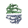
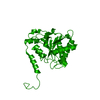
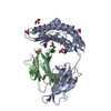
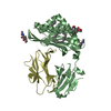
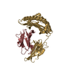
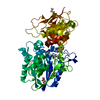
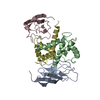
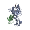
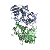
 PRION / PRION REGULATORY DOMAIN / HETEROKARYON INCOMPATIBILITY / PRION- BINDING PROTEIN
 Function and homology information
 Authors
 Citation
 Structure visualization
 Downloads & links
 Other downloads
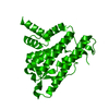

 PDBj
PDBj


 Assembly
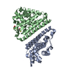


 Mass: 154.251 Da / Num. of mol.: 1 / Source method: obtained synthetically / Formula: C4H10O2S2
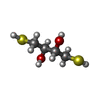
 Mass: 154.251 Da / Num. of mol.: 1 / Source method: obtained synthetically / Formula: C4H10O2S2
 Mass: 18.015 Da / Num. of mol.: 198 / Source method: isolated from a natural source / Formula: H2O
 Sample preparation
 / Beamline: X06SA / Wavelength: 1
  Processing