 Movie
Movie Controller


+ Open data

- Basic information
| Entry | Database: PDB / ID: 2wpk | ||||||
|---|---|---|---|---|---|---|---|
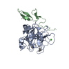
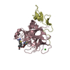
| Title | factor IXa superactive triple mutant, ethylene glycol-soaked | ||||||
 Components Components |
| ||||||
 Keywords Keywords |  BLOOD CLOTTING / HYDROLASE / HEMOSTASIS / HEMOPHILIA BLOOD CLOTTING / HYDROLASE / HEMOSTASIS / HEMOPHILIA | ||||||
| Function / homology |  Function and homology information Function and homology informationDefective F9 secretion / Defective gamma-carboxylation of F9 / coagulation factor IXa / Defective F9 activation / Defective factor IX causes thrombophilia / Defective cofactor function of FVIIIa variant / Defective F9 variant does not activate FX / zymogen activation / Extrinsic Pathway of Fibrin Clot Formation / Protein hydroxylation ...Defective F9 secretion / Defective gamma-carboxylation of F9 / coagulation factor IXa / Defective F9 activation / Defective factor IX causes thrombophilia / Defective cofactor function of FVIIIa variant / Defective F9 variant does not activate FX / zymogen activation / Extrinsic Pathway of Fibrin Clot Formation / Protein hydroxylation / Gamma-carboxylation of protein precursors / Transport of gamma-carboxylated protein precursors from the endoplasmic reticulum to the Golgi apparatus / Removal of aminoterminal propeptides from gamma-carboxylated proteins / Intrinsic Pathway of Fibrin Clot Formation / Golgi lumen / blood coagulation / collagen-containing extracellular matrix / endopeptidase activity / endoplasmic reticulum lumen / serine-type endopeptidase activity / calcium ion binding / proteolysis / extracellular space / extracellular exosome / extracellular region / metal ion binding / plasma membraneSimilarity search - Function | ||||||
| Biological species |  HOMO SAPIENS (human) HOMO SAPIENS (human)SYNTHETIC CONSTRUCT (others) | ||||||
| Method | X-RAY DIFFRACTION / MOLECULAR REPLACEMENT / Resolution: 2.21 Å | ||||||
 Authors Authors | Zogg, T. / Brandstetter, H. | ||||||
 Citation Citation | Journal: Structure / Year: 2009 Title: Structural Basis of the Cofactor- and Substrate-Assisted Activation of Human Coagulation Factor Ixa Authors: Zogg, T. / Brandstetter, H. | ||||||
| History |
| ||||||
| Remark 700 | SHEET DETERMINATION METHOD: DSSP THE SHEETS PRESENTED AS "SA" IN EACH CHAIN ON SHEET RECORDS BELOW ... SHEET DETERMINATION METHOD: DSSP THE SHEETS PRESENTED AS "SA" IN EACH CHAIN ON SHEET RECORDS BELOW IS ACTUALLY AN 7-STRANDED BARREL THIS IS REPRESENTED BY A 8-STRANDED SHEET IN WHICH THE FIRST AND LAST STRANDS ARE IDENTICAL. THE SHEETS PRESENTED AS "SB" IN EACH CHAIN ON SHEET RECORDS BELOW IS ACTUALLY AN 6-STRANDED BARREL THIS IS REPRESENTED BY A 7-STRANDED SHEET IN WHICH THE FIRST AND LAST STRANDS ARE IDENTICAL. |
- Structure visualization
| Structure viewer | Molecule: MolmilJmol/JSmol |
|---|
- Downloads & links
-Download
| PDBx/mmCIF format | 2wpk.cif.gz | 83.6 KB | Display | PDBx/mmCIF format |
|---|---|---|---|---|
| PDB format | pdb2wpk.ent.gz | 59.7 KB | Display | PDB format |
| PDBx/mmJSON format | 2wpk.json.gz | Tree view | PDBx/mmJSON format | |
| Others |  Other downloads Other downloads |
-Validation report
| Arichive directory | https://data.pdbj.org/pub/pdb/validation_reports/wp/2wpkftp://data.pdbj.org/pub/pdb/validation_reports/wp/2wpk | HTTPS FTP |
|---|
-Related structure data
| Related structure data |  2wphSC  2wpiC  2wpjC  2wplC  2wpmC C: citing same article ( S: Starting model for refinement |
|---|---|
| Similar structure data |


-Links
 PDBj
PDBj


- Assembly
| Deposited unit | 
| ||||||||
|---|---|---|---|---|---|---|---|---|---|
| 1 |
| ||||||||
| Unit cell |
|
-Components
-COAGULATION FACTOR IXA ... , 2 types, 2 molecules ES
| #1: Protein | Factor IX / HUMAN COAGULATION FACTOR IXA / CHRISTMAS FACTOR / PLASMA THROMBOPLASTIN COMPONENT / PTC Mass: 6496.395 Da / Num. of mol.: 1 / Fragment: EGF2 DOMAIN, RESIDUES 133-191 Source method: isolated from a genetically manipulated source Source: (gene. exp.) HOMO SAPIENS (human) / Plasmid: PET-FIXA-3X / Production host:  ESCHERICHIA COLI (E. coli) / Strain (production host): BL21(DE3) / References: UniProt: P00740, coagulation factor IXa ESCHERICHIA COLI (E. coli) / Strain (production host): BL21(DE3) / References: UniProt: P00740, coagulation factor IXa |
|---|---|
| #3: Protein | Factor IX / HUMAN COAGULATION FACTOR IXA / CHRISTMAS FACTOR / PLASMA THROMBOPLASTIN COMPONENT / PTC Mass: 26084.672 Da / Num. of mol.: 1 / Fragment: CATALYTIC DOMAIN, RESIDUES 227-461 / Mutation: YES Source method: isolated from a genetically manipulated source Source: (gene. exp.) HOMO SAPIENS (human) / Plasmid: PET-FIXA-3X / Production host: ESCHERICHIA COLI (E. coli) / Strain (production host): BL21(DE3) / References: UniProt: P00740, coagulation factor IXa |
-Protein/peptide , 1 types, 1 molecules L
| #2: Protein/peptide |   Type: Peptide-like / Class: Thrombin inhibitor / Mass: 419.498 Da / Num. of mol.: 1 / Source method: obtained synthetically / Source: (synth.) SYNTHETIC CONSTRUCT (others) / References: BIVALIRUDIN C-terminus fragment Type: Peptide-like / Class: Thrombin inhibitor / Mass: 419.498 Da / Num. of mol.: 1 / Source method: obtained synthetically / Source: (synth.) SYNTHETIC CONSTRUCT (others) / References: BIVALIRUDIN C-terminus fragment |
|---|
-Non-polymers , 3 types, 315 molecules 


| #4: Chemical | ChemComp-CA /  Mass: 40.078 Da / Num. of mol.: 1 / Source method: obtained synthetically / Formula: Ca Mass: 40.078 Da / Num. of mol.: 1 / Source method: obtained synthetically / Formula: Ca |
|---|---|
| #5: Chemical | ChemComp-EDO / Ethylene glycol Mass: 62.068 Da / Num. of mol.: 1 / Source method: obtained synthetically / Formula: C2H6O2 Mass: 62.068 Da / Num. of mol.: 1 / Source method: obtained synthetically / Formula: C2H6O2 |
| #6: Water | ChemComp-HOH / WaterMass: 18.015 Da / Num. of mol.: 313 / Source method: isolated from a natural source / Formula: H2O |
-Experimental details
-Experiment
| Experiment | Method: X-RAY DIFFRACTION / Number of used crystals: 1 |
|---|
- Sample preparation
| Crystal | Density Matthews: 2.14 Å3/Da / Density % sol: 42.7 % / Description: NONE |
|---|---|
| Crystal grow | pH: 6.5 / Details: 22 % PEG 3000 100 MM BIS/TRIS PH 6.85 |
-Data collection
| Diffraction | Mean temperature: 100 K |
|---|---|
| Diffraction source | Source: ROTATING ANODE / Type: BRUKER AXS MICROSTAR / Wavelength: 1.5418 |
| Detector | Type: MARRESEARCH / Detector: IMAGE PLATE / Date: Feb 20, 2008 / Details: MIRRORS |
| Radiation | Protocol: SINGLE WAVELENGTH / Monochromatic (M) / Laue (L): M / Scattering type: x-ray |
| Radiation wavelength | Wavelength: 1.5418 Å / Relative weight: 1 |
| Reflection | Resolution: 2.21→21.6 Å / Num. obs: 14747 / % possible obs: 99.7 % / Observed criterion σ(I): 0 / Redundancy: 6.8 % / Rmerge(I) obs: 0.07 / Net I/σ(I): 22.9 |
| Reflection shell | Resolution: 2.21→2.3 Å / Redundancy: 6.6 % / Rmerge(I) obs: 0.26 / Mean I/σ(I) obs: 6.7 / % possible all: 86.6 |
- Processing
| Software |
| ||||||||||||||||||||||||||||||||||||||||||||||||||||||||||||
|---|---|---|---|---|---|---|---|---|---|---|---|---|---|---|---|---|---|---|---|---|---|---|---|---|---|---|---|---|---|---|---|---|---|---|---|---|---|---|---|---|---|---|---|---|---|---|---|---|---|---|---|---|---|---|---|---|---|---|---|---|---|
| Refinement | Method to determine structure: MOLECULAR REPLACEMENT Starting model: PDB ENTRY 2WPH Resolution: 2.21→21.62 Å / Data cutoff high absF: 10000 / Cross valid method: THROUGHOUT / σ(F): 0 / Stereochemistry target values: MAXIMUM LIKELIHOOD
| ||||||||||||||||||||||||||||||||||||||||||||||||||||||||||||
| Solvent computation | Bsol: 65.68 Å2 / ksol: 0.3 e/Å3 | ||||||||||||||||||||||||||||||||||||||||||||||||||||||||||||
| Displacement parameters |
| ||||||||||||||||||||||||||||||||||||||||||||||||||||||||||||
| Refinement step | Cycle: LAST / Resolution: 2.21→21.62 Å
| ||||||||||||||||||||||||||||||||||||||||||||||||||||||||||||
| Refine LS restraints |
| ||||||||||||||||||||||||||||||||||||||||||||||||||||||||||||
| LS refinement shell | Resolution: 2.21→2.3 Å / Total num. of bins used: 10 | ||||||||||||||||||||||||||||||||||||||||||||||||||||||||||||
| Xplor file |
|
