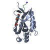
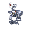
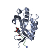
- PDB-2wny: Structure of Mth689, a DUF54 protein from Methanothermobacter the... -
+
Open data
ID or keywords:
Loading...
-
Basic information
Entry
Database: PDB / ID: 2wny
Title
Structure of Mth689, a DUF54 protein from Methanothermobacter thermautotrophicus
Components
CONSERVED PROTEIN MTH689Conservation
Keywords
UNKNOWN FUNCTION / EXOSOME SUPEROPERON
Function / homology
Uncharacterised protein family UPF0201 / RNA binding / 50s Ribosomal Protein L5; Chain: A, / Ribosomal protein L5 / Ribosomal protein L5 domain superfamily / 2-Layer Sandwich / Alpha Beta / NICKEL (II) ION / Conserved protein
Resolution: 1.95→24.23 Å / Cor.coef. Fo:Fc: 0.958 / Cor.coef. Fo:Fc free: 0.919 / SU B: 9.05 / SU ML: 0.122 / Cross valid method: THROUGHOUT / ESU R: 0.159 / ESU R Free: 0.157 / Stereochemistry target values: MAXIMUM LIKELIHOOD Details: HYDROGENS HAVE BEEN ADDED IN THE RIDING POSITIONS. ATOM RECORD CONTAINS SUM OF TLS AND RESIDUAL B FACTORS. ANISOU RECORD CONTAINS SUM OF TLS AND RESIDUAL U FACTORS.
Rfactor
Num. reflection
% reflection
Selection details
Rfree
0.24705
515
2.1 %
RANDOM
Rwork
0.19104
-
-
-
obs
0.19227
23631
99.98 %
-
Solvent computation
Ion probe radii: 0.8 Å / Shrinkage radii: 0.8 Å / VDW probe radii: 1.4 Å / Solvent model: BABINET MODEL WITH MASK
 Movie
Movie Controller

 Yorodumi
 Open data
 Basic information
 Components
 Conservation
  Keywords
 Function and homology information

 Authors
 Citation
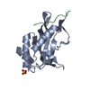
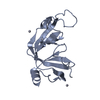
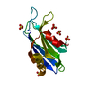
 Structure visualization
 Downloads & links
 Other downloads
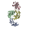


 PDBj
PDBj Assembly
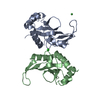


 Mass: 58.693 Da / Num. of mol.: 1 / Source method: obtained synthetically / Formula: Ni
 Mass: 35.453 Da / Num. of mol.: 1 / Source method: obtained synthetically / Formula: Cl
 Mass: 18.015 Da / Num. of mol.: 158 / Source method: isolated from a natural source / Formula: H2O
 Sample preparation
 / Beamline: ID29 / Wavelength: 0.9762
  Processing