 Movie
Movie Controller

[English] 日本語
 Yorodumi
- PDB-2wia: Crystal Structures of the N-terminal Intracellular Domain of FeoB... -
+ Open data

- Basic information
| Entry | Database: PDB / ID: 2wia | ||||||
|---|---|---|---|---|---|---|---|
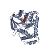
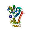
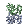
| Title | Crystal Structures of the N-terminal Intracellular Domain of FeoB from Klebsiella Pneumoniae in Apo Form | ||||||
 Components Components | FERROUS IRON TRANSPORT PROTEIN B | ||||||
 Keywords Keywords | METAL TRANSPORT /  SIGNAL TRANSDUCTION / FERROUS IRON TRANSPORT / MEMBRANE PROTEIN / G PROTEIN SIGNAL TRANSDUCTION / FERROUS IRON TRANSPORT / MEMBRANE PROTEIN / G PROTEIN | ||||||
| Function / homology |  Function and homology information Function and homology informationferrous iron transmembrane transporter activity / GTP binding / metal ion binding / plasma membraneSimilarity search - Function | ||||||
| Biological species |  KLEBSIELLA PNEUMONIAE (bacteria) KLEBSIELLA PNEUMONIAE (bacteria) | ||||||
| Method | X-RAY DIFFRACTION / SYNCHROTRON / MOLECULAR REPLACEMENT / Resolution: 2.45 Å | ||||||
 Authors Authors | Hung, K.-W. / Chang, Y.-W. / Chen, J.-H. / Chen, Y.-C. / Sun, Y.-J. / Hsiao, C.-D. / Huang, T.-H. | ||||||
 Citation Citation | Journal: J.Struct.Biol. / Year: 2010 Title: Structural Fold, Conservation and Fe(II) Binding of the Intracellular Domain of Prokaryote Feob. Authors: Hung, K.-W. / Chang, Y.-W. / Eng, E.T. / Chen, J.-H. / Chen, Y.-C. / Sun, Y.-J. / Hsiao, C.-D. / Dong, G. / Spasov, K.A. / Unger, V.M. / Huang, T.-H. | ||||||
| History |
| ||||||
| Remark 650 | HELIX DETERMINATION METHOD: AUTHOR PROVIDED. |
- Structure visualization
| Structure viewer | Molecule: MolmilJmol/JSmol |
|---|
- Downloads & links
-Download
| PDBx/mmCIF format | 2wia.cif.gz | 106.2 KB | Display | PDBx/mmCIF format |
|---|---|---|---|---|
| PDB format | pdb2wia.ent.gz | 85.5 KB | Display | PDB format |
| PDBx/mmJSON format | 2wia.json.gz | Tree view | PDBx/mmJSON format | |
| Others |  Other downloads Other downloads |
-Validation report
| Arichive directory | https://data.pdbj.org/pub/pdb/validation_reports/wi/2wiaftp://data.pdbj.org/pub/pdb/validation_reports/wi/2wia | HTTPS FTP |
|---|
-Related structure data
-Links
 PDBj
PDBj
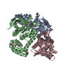
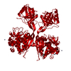
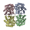
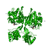
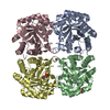
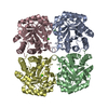
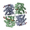
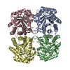
- Assembly
| Deposited unit | 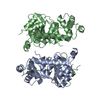
| |||||||||
|---|---|---|---|---|---|---|---|---|---|---|
| 1 | 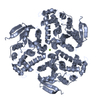
| |||||||||
| 2 | 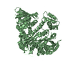
| |||||||||
| 3 | 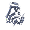
| |||||||||
| 4 | 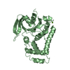
| |||||||||
| Unit cell |
| |||||||||
| Components on special symmetry positions |
|
-Components
| #1: Protein | Mass: 29133.160 Da / Num. of mol.: 2 / Fragment: N-TERMINAL INTRACELLULAR DOMAIN, RESIDUES 1-267 Source method: isolated from a genetically manipulated source Source: (gene. exp.) KLEBSIELLA PNEUMONIAE (bacteria) / Production host: ESCHERICHIA COLI (E. coli) / Strain (production host): BL21(DE3) / References: UniProt: A6TF32#2: Chemical |   Mass: 24.305 Da / Num. of mol.: 2 / Source method: obtained synthetically / Formula: Mg Mass: 24.305 Da / Num. of mol.: 2 / Source method: obtained synthetically / Formula: Mg#3: Water | ChemComp-HOH / | Water Mass: 18.015 Da / Num. of mol.: 198 / Source method: isolated from a natural source / Formula: H2O Mass: 18.015 Da / Num. of mol.: 198 / Source method: isolated from a natural source / Formula: H2O |
|---|
-Experimental details
-Experiment
| Experiment | Method: X-RAY DIFFRACTION |
|---|
- Sample preparation
| Crystal | Density Matthews: 2.09 Å3/Da / Density % sol: 41.2 % / Description: NONE |
|---|---|
| Crystal grow | Details: 100 MM TRIS-HCL PH 8.5, 30% PEG4000, 200 MM MGCL2 |
-Data collection
| Diffraction | Mean temperature: 100 K |
|---|---|
| Diffraction source | Source: SYNCHROTRON / Site: NSRRC  / Beamline: BL13B1 / Wavelength: 1 / Beamline: BL13B1 / Wavelength: 1 |
| Detector | Type: ADSC CCD / Detector: CCD |
| Radiation | Protocol: SINGLE WAVELENGTH / Monochromatic (M) / Laue (L): M / Scattering type: x-ray |
| Radiation wavelength | Wavelength: 1 Å / Relative weight: 1 |
| Reflection | Resolution: 2.45→25.13 Å / Num. obs: 18109 / % possible obs: 99.7 % / Observed criterion σ(I): 2 / Redundancy: 4.6 % / Biso Wilson estimate: 19 Å2 / Rmerge(I) obs: 0.1 / Net I/σ(I): 18.9 |
| Reflection shell | Resolution: 2.45→2.54 Å / Redundancy: 4.7 % / Rmerge(I) obs: 0.28 / Mean I/σ(I) obs: 6.1 / % possible all: 100 |
- Processing
| Software |
| ||||||||||||||||||||||||||||||||||||||||||||||||||||||||||||||||||||||||||||||||
|---|---|---|---|---|---|---|---|---|---|---|---|---|---|---|---|---|---|---|---|---|---|---|---|---|---|---|---|---|---|---|---|---|---|---|---|---|---|---|---|---|---|---|---|---|---|---|---|---|---|---|---|---|---|---|---|---|---|---|---|---|---|---|---|---|---|---|---|---|---|---|---|---|---|---|---|---|---|---|---|---|---|
| Refinement | Method to determine structure: MOLECULAR REPLACEMENT Starting model: STRUCTURE OF FEOB IN GMPPNP STATE Resolution: 2.45→25.13 Å / Rfactor Rfree error: 0.008 / Data cutoff high absF: 59095.74 / Data cutoff low absF: 0 / Isotropic thermal model: RESTRAINED / Cross valid method: THROUGHOUT / σ(F): 2 Details: BULK SOLVENT MODEL USED SIDE CHAIN OF RESIDUES K3, Q51, Q69, T70, E74, E126, K127, Q128, Q129, R131, R152, E173, L187, D224, L226, D227, I228, L233, S234, D235, E236, L242 IN CHAIN A, AND ...Details: BULK SOLVENT MODEL USED SIDE CHAIN OF RESIDUES K3, Q51, Q69, T70, E74, E126, K127, Q128, Q129, R131, R152, E173, L187, D224, L226, D227, I228, L233, S234, D235, E236, L242 IN CHAIN A, AND K3, Q51, T70, E74, I124, E126, K127, Q128, R131, R152, E173, L187, Q190, Q191, S193, Q195, I196, I213, Y214, R216, K225, L226, D227, L233, D235, I237, D238, L242 IN CHAIN B ARE DISORDERED. WHOLE RESIDUE OF T65 TO S68, V149 TO T151 IN CHAIN A, AND T65 TO Q69, V149 TO T151 IN CHAIN B ARE DISORDERED.
| ||||||||||||||||||||||||||||||||||||||||||||||||||||||||||||||||||||||||||||||||
| Solvent computation | Solvent model: FLAT MODEL / Bsol: 38.1939 Å2 / ksol: 0.35 e/Å3 | ||||||||||||||||||||||||||||||||||||||||||||||||||||||||||||||||||||||||||||||||
| Displacement parameters | Biso mean: 30 Å2
| ||||||||||||||||||||||||||||||||||||||||||||||||||||||||||||||||||||||||||||||||
| Refine analyze |
| ||||||||||||||||||||||||||||||||||||||||||||||||||||||||||||||||||||||||||||||||
| Refinement step | Cycle: LAST / Resolution: 2.45→25.13 Å
| ||||||||||||||||||||||||||||||||||||||||||||||||||||||||||||||||||||||||||||||||
| Refine LS restraints |
| ||||||||||||||||||||||||||||||||||||||||||||||||||||||||||||||||||||||||||||||||
| Refine LS restraints NCS | NCS model details: NONE | ||||||||||||||||||||||||||||||||||||||||||||||||||||||||||||||||||||||||||||||||
| LS refinement shell | Resolution: 2.45→2.6 Å / Rfactor Rfree error: 0.017 / Total num. of bins used: 6
| ||||||||||||||||||||||||||||||||||||||||||||||||||||||||||||||||||||||||||||||||
| Xplor file |
|
