 Movie
Movie Controller


+ Open data

- Basic information
| Entry | Database: PDB / ID: 2wfk | ||||||
|---|---|---|---|---|---|---|---|
| Title | Calcium bound LipL32 | ||||||
 Components Components | LIPL32 | ||||||
 Keywords Keywords | METAL BINDING PROTEIN /  FIBRONECTIN / LIPOPROTEIN / CALCIUM BINDING / EXTRACELLULAR MATRIX OUTER MEMBRANE PROTEIN / CA2+-BOUND FIBRONECTIN / LIPOPROTEIN / CALCIUM BINDING / EXTRACELLULAR MATRIX OUTER MEMBRANE PROTEIN / CA2+-BOUND | ||||||
| Function / homology | Surface lipoprotein of Spirochaetales order / Surface lipoprotein of Spirochaetales order / metal ion binding / LipL32 Function and homology information Function and homology information | ||||||
| Biological species |  LEPTOSPIRA SANTAROSAI SEROVAR SHERMANI (bacteria) LEPTOSPIRA SANTAROSAI SEROVAR SHERMANI (bacteria) | ||||||
| Method | X-RAY DIFFRACTION / SYNCHROTRON / MAD / Resolution: 2.3 Å | ||||||
 Authors Authors | Tung, J.-Y. / Yang, C.-W. / Sun, Y.-J. | ||||||
 Citation Citation | Journal: J.Biol.Chem. / Year: 2010 Title: Calcium Binds to Lipl32, a Lipoprotein from Pathogenic Leptospira, and Modulates Fibronectin Binding. Authors: Tung, J.-Y. / Yang, C.-W. / Chou, S. / Lin, C. / Sun, Y.-J. | ||||||
| History |
| ||||||
| Remark 700 | SHEET THE SHEET STRUCTURE OF THIS MOLECULE IS BIFURCATED. IN ORDER TO REPRESENT THIS FEATURE IN ... SHEET THE SHEET STRUCTURE OF THIS MOLECULE IS BIFURCATED. IN ORDER TO REPRESENT THIS FEATURE IN THE SHEET RECORDS BELOW, TWO SHEETS ARE DEFINED. |
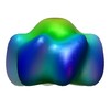
- Structure visualization
| Structure viewer | Molecule: MolmilJmol/JSmol |
|---|
- Downloads & links
-Download
| PDBx/mmCIF format | 2wfk.cif.gz | 486.3 KB | Display | PDBx/mmCIF format |
|---|---|---|---|---|
| PDB format | pdb2wfk.ent.gz | 415.2 KB | Display | PDB format |
| PDBx/mmJSON format | 2wfk.json.gz | Tree view | PDBx/mmJSON format | |
| Others |  Other downloads Other downloads |
-Validation report
| Arichive directory | https://data.pdbj.org/pub/pdb/validation_reports/wf/2wfkftp://data.pdbj.org/pub/pdb/validation_reports/wf/2wfk | HTTPS FTP |
|---|
-Related structure data
| Similar structure data |
|---|


-Links
 PDBj
PDBj- Assembly
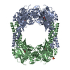
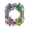
| Deposited unit | 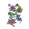
| ||||||||
|---|---|---|---|---|---|---|---|---|---|
| 1 | 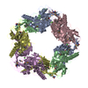
| ||||||||
| Unit cell |
|
-Components
| #1: Protein | Mass: 28793.953 Da / Num. of mol.: 5 / Fragment: RESIDUES 20-266 Source method: isolated from a genetically manipulated source Source: (gene. exp.) LEPTOSPIRA SANTAROSAI SEROVAR SHERMANI (bacteria)Plasmid: PRSETC / Production host: ESCHERICHIA COLI (E. coli) / Strain (production host): BL21(DE3) PLYS / References: UniProt: Q6S9S1#2: Chemical | ChemComp-CA /   Mass: 40.078 Da / Num. of mol.: 5 / Source method: obtained synthetically / Formula: Ca Mass: 40.078 Da / Num. of mol.: 5 / Source method: obtained synthetically / Formula: Ca#3: Water | ChemComp-HOH / | Water Mass: 18.015 Da / Num. of mol.: 528 / Source method: isolated from a natural source / Formula: H2O Mass: 18.015 Da / Num. of mol.: 528 / Source method: isolated from a natural source / Formula: H2O |
|---|
-Experimental details
-Experiment
| Experiment | Method: X-RAY DIFFRACTION |
|---|
- Sample preparation
| Crystal | Density Matthews: 2.94 Å3/Da / Density % sol: 55.42 % / Description: NONE |
|---|---|
| Crystal grow | Details: TACSIMATE, PH7.0 |
-Data collection
| Diffraction | Mean temperature: 287 K |
|---|---|
| Diffraction source | Source: SYNCHROTRON / Site: SPring-8  / Beamline: BL12B2 / Wavelength: 1 / Beamline: BL12B2 / Wavelength: 1 |
| Radiation | Protocol: MAD / Monochromatic (M) / Laue (L): M / Scattering type: x-ray |
| Radiation wavelength | Wavelength: 1 Å / Relative weight: 1 |
| Reflection | Resolution: 2.3→30 Å / Num. obs: 69717 / % possible obs: 99.7 % / Observed criterion σ(I): 2 / Redundancy: 13.8 % / Biso Wilson estimate: 44.85 Å2 / Rmerge(I) obs: 0.05 / Net I/σ(I): 47.2 |
| Reflection shell | Resolution: 2.3→2.38 Å / Redundancy: 14.1 % / Rmerge(I) obs: 0.42 / Mean I/σ(I) obs: 6.7 / % possible all: 100 |
- Processing
| Software |
| ||||||||||||||||||||||||||||||||||||||||||||||||||||||||||||||||||||||||||||||||||||||||||||||||||||||||||||||||||||||||||||||||||||||||||||||||||||||||||||||||||||||||||||||||||||||
|---|---|---|---|---|---|---|---|---|---|---|---|---|---|---|---|---|---|---|---|---|---|---|---|---|---|---|---|---|---|---|---|---|---|---|---|---|---|---|---|---|---|---|---|---|---|---|---|---|---|---|---|---|---|---|---|---|---|---|---|---|---|---|---|---|---|---|---|---|---|---|---|---|---|---|---|---|---|---|---|---|---|---|---|---|---|---|---|---|---|---|---|---|---|---|---|---|---|---|---|---|---|---|---|---|---|---|---|---|---|---|---|---|---|---|---|---|---|---|---|---|---|---|---|---|---|---|---|---|---|---|---|---|---|---|---|---|---|---|---|---|---|---|---|---|---|---|---|---|---|---|---|---|---|---|---|---|---|---|---|---|---|---|---|---|---|---|---|---|---|---|---|---|---|---|---|---|---|---|---|---|---|---|---|
| Refinement | Method to determine structure: MAD Starting model: NONE Resolution: 2.3→27.259 Å / SU ML: 0.36 / σ(F): 2 / Phase error: 26.81 / Stereochemistry target values: ML Details: DISORDERED REGIONS WERE MISSING FROM RESIUDE (B PRO 168 TO B THR 182), (C ILE 167 TO C ASN 179), (D ILE 165 TO D ILE 180)
| ||||||||||||||||||||||||||||||||||||||||||||||||||||||||||||||||||||||||||||||||||||||||||||||||||||||||||||||||||||||||||||||||||||||||||||||||||||||||||||||||||||||||||||||||||||||
| Solvent computation | Shrinkage radii: 0.9 Å / VDW probe radii: 1.11 Å / Solvent model: FLAT BULK SOLVENT MODEL / Bsol: 44.328 Å2 / ksol: 0.34 e/Å3 | ||||||||||||||||||||||||||||||||||||||||||||||||||||||||||||||||||||||||||||||||||||||||||||||||||||||||||||||||||||||||||||||||||||||||||||||||||||||||||||||||||||||||||||||||||||||
| Displacement parameters | Biso mean: 49.5 Å2
| ||||||||||||||||||||||||||||||||||||||||||||||||||||||||||||||||||||||||||||||||||||||||||||||||||||||||||||||||||||||||||||||||||||||||||||||||||||||||||||||||||||||||||||||||||||||
| Refinement step | Cycle: LAST / Resolution: 2.3→27.259 Å
| ||||||||||||||||||||||||||||||||||||||||||||||||||||||||||||||||||||||||||||||||||||||||||||||||||||||||||||||||||||||||||||||||||||||||||||||||||||||||||||||||||||||||||||||||||||||
| Refine LS restraints |
| ||||||||||||||||||||||||||||||||||||||||||||||||||||||||||||||||||||||||||||||||||||||||||||||||||||||||||||||||||||||||||||||||||||||||||||||||||||||||||||||||||||||||||||||||||||||
| LS refinement shell |
|
