[English] 日本語
Yorodumi
- PDB-2wfd: Structure of the human cytosolic leucyl-tRNA synthetase editing domain -

+
Open data


ID or keywords:

Loading...

-
Basic information

Entry
Database: PDB / ID: 2wfd
TitleStructure of the human cytosolic leucyl-tRNA synthetase editing domain
ComponentsLEUCYL-TRNA SYNTHETASE, CYTOPLASMIC
KeywordsLIGASE / AMINOACYL-TRNA SYNTHETASE / PHOSPHOPROTEIN / EDITING DOMAIN / NUCLEOTIDE-BINDING / HYDROLYSIS OF MIS-CHARGED TRNAS / PROTEIN BIOSYNTHESIS / LEUCYL-TRNA SYNTHETASE / HUMAN / CYTOPLASM / ATP-BINDING / POLYMORPHISM
Function / homology
Function and homology information


glutamine-tRNA ligase activity / glutaminyl-tRNA aminoacylation / Selenoamino acid metabolism / leucine-tRNA ligase / leucine-tRNA ligase activity / leucyl-tRNA aminoacylation / aminoacyl-tRNA synthetase multienzyme complex / cellular response to leucine starvation / Cytosolic tRNA aminoacylation / cellular response to L-leucine ...glutamine-tRNA ligase activity / glutaminyl-tRNA aminoacylation / Selenoamino acid metabolism / leucine-tRNA ligase / leucine-tRNA ligase activity / leucyl-tRNA aminoacylation / aminoacyl-tRNA synthetase multienzyme complex / cellular response to leucine starvation / Cytosolic tRNA aminoacylation / cellular response to L-leucine / tRNA aminoacylation for protein translation / aminoacyl-tRNA editing activity / regulation of cell size / positive regulation of TOR signaling / endomembrane system / positive regulation of TORC1 signaling / cellular response to amino acid starvation / GTPase activator activity / cellular response to amino acid stimulus / positive regulation of GTPase activity / lysosome / nuclear body / endoplasmic reticulum / ATP binding / cytosol / cytoplasm
Similarity search - Function
Leucyl-tRNA synthetase, class Ia, archaeal/eukaryotic cytosolic / Isoleucyl-tRNA Synthetase; domain 2 / Valyl/Leucyl/Isoleucyl-tRNA synthetase, editing domain / Aminoacyl-tRNA synthetase, class Ia / tRNA synthetases class I (I, L, M and V) / Valyl/Leucyl/Isoleucyl-tRNA synthetase, editing domain / Methionyl/Valyl/Leucyl/Isoleucyl-tRNA synthetase, anticodon-binding / Anticodon-binding domain of tRNA ligase / Aminoacyl-tRNA synthetase, class Ia, anticodon-binding / Aminoacyl-tRNA synthetase, class I, conserved site ...Leucyl-tRNA synthetase, class Ia, archaeal/eukaryotic cytosolic / Isoleucyl-tRNA Synthetase; domain 2 / Valyl/Leucyl/Isoleucyl-tRNA synthetase, editing domain / Aminoacyl-tRNA synthetase, class Ia / tRNA synthetases class I (I, L, M and V) / Valyl/Leucyl/Isoleucyl-tRNA synthetase, editing domain / Methionyl/Valyl/Leucyl/Isoleucyl-tRNA synthetase, anticodon-binding / Anticodon-binding domain of tRNA ligase / Aminoacyl-tRNA synthetase, class Ia, anticodon-binding / Aminoacyl-tRNA synthetase, class I, conserved site / Aminoacyl-transfer RNA synthetases class-I signature. / Rossmann-like alpha/beta/alpha sandwich fold / Alpha-Beta Complex / Alpha Beta
Similarity search - Domain/homology
Leucine--tRNA ligase, cytoplasmic
Similarity search - Component
Biological speciesHOMO SAPIENS (human)
MethodX-RAY DIFFRACTION / SYNCHROTRON / SAD / Resolution: 3.25 Å
AuthorsSeiradake, E. / Mao, W. / Hernandez, V. / Baker, S.J. / Plattner, J.J. / Alley, M.R.K. / Cusack, S.
CitationJournal: J.Mol.Biol. / Year: 2009
Title: Crystal Structures of the Human and Fungal Cytosolic Leucyl-tRNA Synthetase Editing Domains: A Structural Basis for the Rational Design of Antifungal Benzoxaboroles.
Authors: Seiradake, E. / Mao, W. / Hernandez, V. / Baker, S.J. / Plattner, J.J. / Alley, M.R.K. / Cusack, S.
History
DepositionApr 3, 2009Deposition site: PDBE / Processing site: PDBE
Revision 1.0May 19, 2009Provider: repository / Type: Initial release
Revision 1.1May 8, 2011Group: Version format compliance
Revision 1.2Jul 13, 2011Group: Version format compliance
Remark 700 
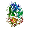
SHEET THE SHEET STRUCTURE OF THIS MOLECULE IS BIFURCATED. IN ORDER TO REPRESENT THIS FEATURE IN ... SHEET THE SHEET STRUCTURE OF THIS MOLECULE IS BIFURCATED. IN ORDER TO REPRESENT THIS FEATURE IN THE SHEET RECORDS BELOW, TWO SHEETS ARE DEFINED.

-
Structure visualization

Structure viewerMolecule:
MolmilJmol/JSmol

Downloads & links

-
Assembly

Deposited unit
A: LEUCYL-TRNA SYNTHETASE, CYTOPLASMIC
B: LEUCYL-TRNA SYNTHETASE, CYTOPLASMIC


Theoretical massNumber of molelcules
Total (without water)56,9432
Polymers56,9432
Non-polymers00
Water0
1


  • Idetical with deposited unit
  • defined by author&software
TypeNameSymmetry operationNumber
identity operation1_555x,y,z1
Buried area2700 Å2
ΔGint-18.93 kcal/mol
Surface area21880 Å2
MethodPISA
Unit cell
Length a, b, c (Å)94.540, 94.540, 148.820
Angle α, β, γ (deg.)90.00, 90.00, 120.00
Int Tables number170
Space group name H-MP65
Noncrystallographic symmetry (NCS)NCS domain:
IDEns-IDDetails
11A
21B

NCS domain segments:
Dom-IDComponent-IDEns-IDRefine codeAuth asym-IDAuth seq-ID
1111A258 - 401
2111B258 - 401
1211A425 - 509
2211B425 - 509

-
Components

#1: Protein LEUCYL-TRNA SYNTHETASE, CYTOPLASMIC / / LEUCINE--TRNA LIGASE / LEURS


Mass: 28471.697 Da / Num. of mol.: 2 / Fragment: EDITING OR CP1 DOMAIN, RESIDUES 260-509
Source method: isolated from a genetically manipulated source
Source: (gene. exp.) HOMO SAPIENS (human) / Plasmid: PPROEX HTB / Production host: ESCHERICHIA COLI (E. coli) / Strain (production host): BL21(DE3) / References: UniProt: Q9P2J5, leucine-tRNA ligase
Nonpolymer detailsSELENOMETHIONINE (MSE): PROTEIN WAS SELENOMETHIONINE LABELLED
Sequence detailsEXTRA ALA-MET AT N-TERMINUS FROM CLONING.

-
Experimental details

-
Experiment

ExperimentMethod: X-RAY DIFFRACTION / Number of used crystals: 1

-
Sample preparation

CrystalDensity Matthews: 3.45 Å3/Da / Density % sol: 63.2 % / Description: NONE
Crystal growDetails: 0.2 M SODIUM CITRATE TRIBASIC DIHYDRATE PH 8.3, 20% W/V POLYETHYLENE GLYCOL 3350

-
Data collection

DiffractionMean temperature: 100 K
Diffraction sourceSource: SYNCHROTRON / Site: ESRF / Beamline: ID14-4 / Wavelength: 0.9795
DetectorType: ADSC CCD / Detector: CCD
RadiationProtocol: SINGLE WAVELENGTH / Monochromatic (M) / Laue (L): M / Scattering type: x-ray
Radiation wavelengthWavelength: 0.9795 Å / Relative weight: 1
ReflectionResolution: 3→30 Å / Num. obs: 15008 / % possible obs: 99.8 % / Observed criterion σ(I): 0 / Redundancy: 3.12 % / Rmerge(I) obs: 0.15 / Net I/σ(I): 7.46
Reflection shellResolution: 3.25→3.3 Å / Redundancy: 3.16 % / Rmerge(I) obs: 0.66 / Mean I/σ(I) obs: 1.98 / % possible all: 99.5

-
Processing

Software
NameVersionClassification
REFMAC5.5.0070refinement
XDSdata reduction
XSCALEdata scaling
SHELXDSHARPphasing
RefinementMethod to determine structure: SAD
Starting model: NONE

Resolution: 3.25→29.24 Å / Cor.coef. Fo:Fc: 0.92 / Cor.coef. Fo:Fc free: 0.894 / SU B: 50.954 / SU ML: 0.378 / Cross valid method: THROUGHOUT / ESU R Free: 0.471 / Stereochemistry target values: MAXIMUM LIKELIHOOD
Details: HYDROGENS HAVE BEEN ADDED IN THE RIDING POSITIONS. U VALUES RESIDUAL ONLY
RfactorNum. reflection% reflectionSelection details
Rfree0.24 612 5.2 %RANDOM
Rwork0.202 ---
obs0.204 11198 100 %-
Solvent computationIon probe radii: 0.8 Å / Shrinkage radii: 0.8 Å / VDW probe radii: 1.4 Å / Solvent model: MASK
Refinement stepCycle: LAST / Resolution: 3.25→29.24 Å
ProteinNucleic acidLigandSolventTotal
Num. atoms3922 0 0 0 3922
Refine LS restraints
Refine-IDTypeDev idealDev ideal targetNumber
X-RAY DIFFRACTIONr_bond_refined_d0.010.0223990
X-RAY DIFFRACTIONr_bond_other_d0.0010.022788
X-RAY DIFFRACTIONr_angle_refined_deg1.3182.0045380
X-RAY DIFFRACTIONr_angle_other_deg0.85236886
X-RAY DIFFRACTIONr_dihedral_angle_1_deg6.8165502
X-RAY DIFFRACTIONr_dihedral_angle_2_deg43.27325.526152
X-RAY DIFFRACTIONr_dihedral_angle_3_deg17.80615770
X-RAY DIFFRACTIONr_dihedral_angle_4_deg17.3211514
X-RAY DIFFRACTIONr_chiral_restr0.0690.2614
X-RAY DIFFRACTIONr_gen_planes_refined0.0040.0214318
X-RAY DIFFRACTIONr_gen_planes_other0.0010.02710
X-RAY DIFFRACTIONr_nbd_refined
X-RAY DIFFRACTIONr_nbd_other
X-RAY DIFFRACTIONr_nbtor_refined
X-RAY DIFFRACTIONr_nbtor_other
X-RAY DIFFRACTIONr_xyhbond_nbd_refined
X-RAY DIFFRACTIONr_xyhbond_nbd_other
X-RAY DIFFRACTIONr_metal_ion_refined
X-RAY DIFFRACTIONr_metal_ion_other
X-RAY DIFFRACTIONr_symmetry_vdw_refined
X-RAY DIFFRACTIONr_symmetry_vdw_other
X-RAY DIFFRACTIONr_symmetry_hbond_refined
X-RAY DIFFRACTIONr_symmetry_hbond_other
X-RAY DIFFRACTIONr_symmetry_metal_ion_refined
X-RAY DIFFRACTIONr_symmetry_metal_ion_other
X-RAY DIFFRACTIONr_mcbond_it0.3361.52508
X-RAY DIFFRACTIONr_mcbond_other0.0431.51018
X-RAY DIFFRACTIONr_mcangle_it0.6524064
X-RAY DIFFRACTIONr_mcangle_other
X-RAY DIFFRACTIONr_scbond_it0.94331482
X-RAY DIFFRACTIONr_scbond_other
X-RAY DIFFRACTIONr_scangle_it1.5714.51316
X-RAY DIFFRACTIONr_scangle_other
X-RAY DIFFRACTIONr_long_range_B_refined
X-RAY DIFFRACTIONr_long_range_B_other
X-RAY DIFFRACTIONr_rigid_bond_restr
X-RAY DIFFRACTIONr_sphericity_free
X-RAY DIFFRACTIONr_sphericity_bonded
Refine LS restraints NCS

Ens-ID: 1 / Number: 3019 / Refine-ID: X-RAY DIFFRACTION

Dom-IDAuth asym-IDTypeRms dev position (Å)Weight position
1Atight positional0.030.05
2Btight positional0.030.05
1Atight thermal0.885
2Btight thermal0.885
LS refinement shellResolution: 3.25→3.33 Å / Total num. of bins used: 20
RfactorNum. reflection% reflection
Rfree0.25 46 -
Rwork0.3 812 -
obs--100 %
Refinement TLS params.

Method: refined / Refine-ID: X-RAY DIFFRACTION

IDL112)L122)L132)L222)L232)L332)S11 (Å °)S12 (Å °)S13 (Å °)S21 (Å °)S22 (Å °)S23 (Å °)S31 (Å °)S32 (Å °)S33 (Å °)T112)T122)T132)T222)T232)T332)Origin x (Å)Origin y (Å)Origin z (Å)
14.28932.35310.2891.68410.09951.69870.0355-0.25510.1820.1789-0.02850.0712-0.1952-0.156-0.0069-0.2567-0.03460.0132-0.0849-0.0633-0.23225.249-37.795-15.778
23.95461.5832-0.99271.7456-0.1084.1614-0.07540.51180.0413-0.11610.02610.29950.2456-1.09050.0493-0.2598-0.10870.0193-0.0426-0.04-0.182827.48-37.833-48.714
Refinement TLS group
IDRefine-IDRefine TLS-IDAuth asym-IDAuth seq-ID
1X-RAY DIFFRACTION1A258 - 509
2X-RAY DIFFRACTION2B258 - 509

+
About Yorodumi

-
News

-
Feb 9, 2022. New format data for meta-information of EMDB entries

New format data for meta-information of EMDB entries

  • Version 3 of the EMDB header file is now the official format.
  • The previous official version 1.9 will be removed from the archive.

Related info.:EMDB header

External links:wwPDB to switch to version 3 of the EMDB data model

-
Aug 12, 2020. Covid-19 info

Covid-19 info

URL: https://pdbj.org/emnavi/covid19.php

New page: Covid-19 featured information page in EM Navigator.

Related info.:Covid-19 info / Mar 5, 2020. Novel coronavirus structure data

+
Mar 5, 2020. Novel coronavirus structure data

Novel coronavirus structure data

Related info.:Yorodumi Speices / Aug 12, 2020. Covid-19 info

External links:COVID-19 featured content - PDBj / Molecule of the Month (242):Coronavirus Proteases

+
Jan 31, 2019. EMDB accession codes are about to change! (news from PDBe EMDB page)

EMDB accession codes are about to change! (news from PDBe EMDB page)

  • The allocation of 4 digits for EMDB accession codes will soon come to an end. Whilst these codes will remain in use, new EMDB accession codes will include an additional digit and will expand incrementally as the available range of codes is exhausted. The current 4-digit format prefixed with “EMD-” (i.e. EMD-XXXX) will advance to a 5-digit format (i.e. EMD-XXXXX), and so on. It is currently estimated that the 4-digit codes will be depleted around Spring 2019, at which point the 5-digit format will come into force.
  • The EM Navigator/Yorodumi systems omit the EMD- prefix.

Related info.:Q: What is EMD? / ID/Accession-code notation in Yorodumi/EM Navigator

External links:EMDB Accession Codes are Changing Soon! / Contact to PDBj

+
Jul 12, 2017. Major update of PDB

Major update of PDB

  • wwPDB released updated PDB data conforming to the new PDBx/mmCIF dictionary.
  • This is a major update changing the version number from 4 to 5, and with Remediation, in which all the entries are updated.
  • In this update, many items about electron microscopy experimental information are reorganized (e.g. em_software).
  • Now, EM Navigator and Yorodumi are based on the updated data.

External links:wwPDB Remediation / Enriched Model Files Conforming to OneDep Data Standards Now Available in the PDB FTP Archive

-
Yorodumi

Thousand views of thousand structures

  • Yorodumi is a browser for structure data from EMDB, PDB, SASBDB, etc.
  • This page is also the successor to EM Navigator detail page, and also detail information page/front-end page for Omokage search.
  • The word "yorodu" (or yorozu) is an old Japanese word meaning "ten thousand". "mi" (miru) is to see.

Related info.:EMDB / PDB / SASBDB / Comparison of 3 databanks / Yorodumi Search / Aug 31, 2016. New EM Navigator & Yorodumi / Yorodumi Papers / Jmol/JSmol / Function and homology information / Changes in new EM Navigator and Yorodumi

Read more