 Movie
Movie Controller

[English] 日本語
 Yorodumi
- PDB-2wc9: Crystal structure of the g2p (large terminase) nuclease domain fr... -
+ Open data

- Basic information
| Entry | Database: PDB / ID: 2wc9 | ||||||
|---|---|---|---|---|---|---|---|
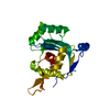
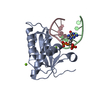
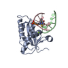
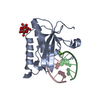
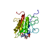
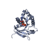
| Title | Crystal structure of the g2p (large terminase) nuclease domain from the bacteriophage SPP1 with bound Mn | ||||||
 Components Components | TERMINASE LARGE SUBUNIT | ||||||
 Keywords Keywords |  VIRAL PROTEIN / DNA TRANSLOCATION VIRAL PROTEIN / DNA TRANSLOCATION | ||||||
| Function / homology |  Function and homology information Function and homology informationviral terminase, large subunit / viral DNA genome packaging / nuclease activity / chromosome organization / Hydrolases; Acting on acid anhydrides; Acting on acid anhydrides to facilitate cellular and subcellular movement / endonuclease activity / Hydrolases; Acting on ester bonds / ATP hydrolysis activity / ATP binding / metal ion bindingSimilarity search - Function | ||||||
| Biological species |  BACILLUS PHAGE SPP1 (virus) BACILLUS PHAGE SPP1 (virus) | ||||||
| Method | X-RAY DIFFRACTION / OTHER / Resolution: 2.5 Å | ||||||
 Authors Authors | Smits, C. / Chechik, M. / Kovalevskiy, O.V. / Shevtsov, M.B. / Foster, A.W. / Alonso, J.C. / Antson, A.A. | ||||||
 Citation Citation | Journal: Embo Rep. / Year: 2009 Title: Structural Basis for the Nuclease Activity of a Bacteriophage Large Terminase. Authors: Smits, C. / Chechik, M. / Kovalevskiy, O.V. / Shevtsov, M.B. / Foster, A.W. / Alonso, J.C. / Antson, A.A. | ||||||
| History |
|
- Structure visualization
| Structure viewer | Molecule: MolmilJmol/JSmol |
|---|
- Downloads & links
-Download
| PDBx/mmCIF format | 2wc9.cif.gz | 45.8 KB | Display | PDBx/mmCIF format |
|---|---|---|---|---|
| PDB format | pdb2wc9.ent.gz | 35.4 KB | Display | PDB format |
| PDBx/mmJSON format | 2wc9.json.gz | Tree view | PDBx/mmJSON format | |
| Others |  Other downloads Other downloads |
-Validation report
| Arichive directory | https://data.pdbj.org/pub/pdb/validation_reports/wc/2wc9ftp://data.pdbj.org/pub/pdb/validation_reports/wc/2wc9 | HTTPS FTP |
|---|
-Related structure data


-Links
 PDBj
PDBj- Assembly
| Deposited unit | 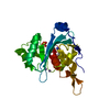
| ||||||||
|---|---|---|---|---|---|---|---|---|---|
| 1 |
| ||||||||
| Unit cell |
|
-Components
| #1: Protein | Mass: 24296.648 Da / Num. of mol.: 1 / Fragment: NUCLEASE DOMAIN, RESIDUES 232-422 Source method: isolated from a genetically manipulated source Source: (gene. exp.) BACILLUS PHAGE SPP1 (virus) / Plasmid: PYM20 / Production host:  ESCHERICHIA COLI (E. coli) / Strain (production host): B834 (DE3) / References: UniProt: P54308 ESCHERICHIA COLI (E. coli) / Strain (production host): B834 (DE3) / References: UniProt: P54308 | ||||
|---|---|---|---|---|---|
| #2: Chemical |   Mass: 54.938 Da / Num. of mol.: 2 / Source method: obtained synthetically / Formula: Mn Mass: 54.938 Da / Num. of mol.: 2 / Source method: obtained synthetically / Formula: Mn#3: Chemical | ChemComp-SO4 / | Sulfate  Mass: 96.063 Da / Num. of mol.: 1 / Source method: obtained synthetically / Formula: SO4 Mass: 96.063 Da / Num. of mol.: 1 / Source method: obtained synthetically / Formula: SO4#4: Water | ChemComp-HOH / | Water Mass: 18.015 Da / Num. of mol.: 24 / Source method: isolated from a natural source / Formula: H2O Mass: 18.015 Da / Num. of mol.: 24 / Source method: isolated from a natural source / Formula: H2O |
-Experimental details
-Experiment
| Experiment | Method: X-RAY DIFFRACTION / Number of used crystals: 1 |
|---|
- Sample preparation
| Crystal | Density Matthews: 2.1 Å3/Da / Density % sol: 42 % / Description: NONE |
|---|---|
| Crystal grow | Details: 80 MM TRIS PH 8.5, 2.3 M LI2SO4 |
-Data collection
| Diffraction | Mean temperature: 120 K |
|---|---|
| Diffraction source | Source: ROTATING ANODE / Type: RIGAKU MICROMAX-007 HF / Wavelength: 1.5418 |
| Detector | Detector: IMAGE PLATE / Date: Sep 20, 2008 |
| Radiation | Protocol: SINGLE WAVELENGTH / Monochromatic (M) / Laue (L): M / Scattering type: x-ray |
| Radiation wavelength | Wavelength: 1.5418 Å / Relative weight: 1 |
| Reflection | Resolution: 2.5→60.4 Å / Num. obs: 7520 / % possible obs: 100 % / Observed criterion σ(I): 2 / Redundancy: 11.8 % / Rmerge(I) obs: 0.05 / Net I/σ(I): 32.4 |
| Reflection shell | Resolution: 2.5→2.64 Å / Redundancy: 11.8 % / Rmerge(I) obs: 0.54 / Mean I/σ(I) obs: 4.4 / % possible all: 100 |
- Processing
| Software |
| ||||||||||||||||||||||||||||||||||||||||||||||||||||||||||||||||||||||||||||||||||||||||||||||||||||||||||||||||||||||||||||||||||||||||||||||||||||||||||||||||||||||||||||||||||||||
|---|---|---|---|---|---|---|---|---|---|---|---|---|---|---|---|---|---|---|---|---|---|---|---|---|---|---|---|---|---|---|---|---|---|---|---|---|---|---|---|---|---|---|---|---|---|---|---|---|---|---|---|---|---|---|---|---|---|---|---|---|---|---|---|---|---|---|---|---|---|---|---|---|---|---|---|---|---|---|---|---|---|---|---|---|---|---|---|---|---|---|---|---|---|---|---|---|---|---|---|---|---|---|---|---|---|---|---|---|---|---|---|---|---|---|---|---|---|---|---|---|---|---|---|---|---|---|---|---|---|---|---|---|---|---|---|---|---|---|---|---|---|---|---|---|---|---|---|---|---|---|---|---|---|---|---|---|---|---|---|---|---|---|---|---|---|---|---|---|---|---|---|---|---|---|---|---|---|---|---|---|---|---|---|
| Refinement | Method to determine structure: OTHER Starting model: NONE Resolution: 2.5→24.6 Å / Cor.coef. Fo:Fc: 0.954 / Cor.coef. Fo:Fc free: 0.924 / SU B: 21.218 / SU ML: 0.211 / Cross valid method: THROUGHOUT / ESU R: 0.546 / ESU R Free: 0.299 / Stereochemistry target values: MAXIMUM LIKELIHOOD / Details: HYDROGENS HAVE BEEN ADDED IN THE RIDING POSITIONS.
| ||||||||||||||||||||||||||||||||||||||||||||||||||||||||||||||||||||||||||||||||||||||||||||||||||||||||||||||||||||||||||||||||||||||||||||||||||||||||||||||||||||||||||||||||||||||
| Solvent computation | Ion probe radii: 0.8 Å / Shrinkage radii: 0.8 Å / VDW probe radii: 1.2 Å / Solvent model: BABINET MODEL WITH MASK | ||||||||||||||||||||||||||||||||||||||||||||||||||||||||||||||||||||||||||||||||||||||||||||||||||||||||||||||||||||||||||||||||||||||||||||||||||||||||||||||||||||||||||||||||||||||
| Displacement parameters | Biso mean: 32.121 Å2
| ||||||||||||||||||||||||||||||||||||||||||||||||||||||||||||||||||||||||||||||||||||||||||||||||||||||||||||||||||||||||||||||||||||||||||||||||||||||||||||||||||||||||||||||||||||||
| Refinement step | Cycle: LAST / Resolution: 2.5→24.6 Å
| ||||||||||||||||||||||||||||||||||||||||||||||||||||||||||||||||||||||||||||||||||||||||||||||||||||||||||||||||||||||||||||||||||||||||||||||||||||||||||||||||||||||||||||||||||||||
| Refine LS restraints |
|