[English] 日本語
Yorodumi
- PDB-2w03: Co-complex Structure of Achromobactin Synthetase Protein D (AcsD)... -

+
Open data


ID or keywords:

Loading...

-
Basic information

Entry
Database: PDB / ID: 2w03
TitleCo-complex Structure of Achromobactin Synthetase Protein D (AcsD) with adenosine, sulfate and citrate from Pectobacterium Chrysanthemi
ComponentsACSD
KeywordsMETAL TRANSPORT / SSPF / ACSD / ACHROMOBACTIN BIOSYNTHESIS / PECTOBACTERIUM CHRYSANTHEMI
Function / homology
Function and homology information


acid-amino acid ligase activity / siderophore biosynthetic process / ATP binding / metal ion binding
Similarity search - Function
AcsD, thumb domain, helical bundle / AcsD, palm domain, helix bundle / N-terminal domain of TfIIb - #450 / AscD, thumb domain, four stranded beta-sheet / Double Stranded RNA Binding Domain - #870 / PvsD/AcsD-like, thumb domain, helical bundle / PvsD/AcsD-like, thumb domain, four stranded beta-sheet / PvsD/AcsD-like, palm domain, helix bundle / Aerobactin siderophore biosynthesis, IucA/IucC, N-terminal / Aerobactin siderophore biosynthesis, IucA/IucC-like ...AcsD, thumb domain, helical bundle / AcsD, palm domain, helix bundle / N-terminal domain of TfIIb - #450 / AscD, thumb domain, four stranded beta-sheet / Double Stranded RNA Binding Domain - #870 / PvsD/AcsD-like, thumb domain, helical bundle / PvsD/AcsD-like, thumb domain, four stranded beta-sheet / PvsD/AcsD-like, palm domain, helix bundle / Aerobactin siderophore biosynthesis, IucA/IucC, N-terminal / Aerobactin siderophore biosynthesis, IucA/IucC-like / IucA / IucC family / Ferric iron reductase FhuF domain / Ferric iron reductase FhuF-like transporter / Endonuclease III; domain 1 / N-terminal domain of TfIIb / Double Stranded RNA Binding Domain / Single Sheet / SH3 type barrels. / DNA polymerase; domain 1 / Roll / 2-Layer Sandwich / Orthogonal Bundle / Mainly Beta / Mainly Alpha / Alpha Beta
Similarity search - Domain/homology
ADENOSINE / CITRIC ACID / AcsD
Similarity search - Component
Biological speciesERWINIA CHRYSANTHEMI (bacteria)
MethodX-RAY DIFFRACTION / SYNCHROTRON / MOLECULAR REPLACEMENT / Resolution: 2.95 Å
AuthorsSchmelz, S. / McMahon, S.A. / Kadi, N. / Song, L. / Oves-Costales, D. / Oke, M. / Liu, H. / Johnson, K.A. / Carter, L. / White, M.F. ...Schmelz, S. / McMahon, S.A. / Kadi, N. / Song, L. / Oves-Costales, D. / Oke, M. / Liu, H. / Johnson, K.A. / Carter, L. / White, M.F. / Challis, G.L. / Naismith, J.H.
CitationJournal: Nat.Chem.Biol. / Year: 2009
Title: Acsd Catalyzes Enantioselective Citrate Desymmetrization in Siderophore Biosynthesis
Authors: Schmelz, S. / Kadi, N. / Mcmahon, S.A. / Song, L. / Oves-Costales, D. / Oke, M. / Liu, H. / Johnson, K.A. / Carter, L. / Botting, C.H. / White, M.F. / Challis, G.L. / Naismith, J.H.
History
DepositionAug 8, 2008Deposition site: PDBE / Processing site: PDBE
Revision 1.0Jan 13, 2009Provider: repository / Type: Initial release
Revision 1.1May 7, 2011Group: Version format compliance
Revision 1.2Jul 13, 2011Group: Version format compliance
Revision 1.3May 8, 2019Group: Data collection / Experimental preparation / Other
Category: database_PDB_rev / database_PDB_rev_record ...database_PDB_rev / database_PDB_rev_record / exptl_crystal_grow / pdbx_database_proc / pdbx_database_status
Item: _exptl_crystal_grow.method / _pdbx_database_status.recvd_author_approval

-
Structure visualization

Structure viewerMolecule:
MolmilJmol/JSmol

Downloads & links

-
Assembly

Deposited unit
A: ACSD
B: ACSD
hetero molecules


Theoretical massNumber of molelcules
Total (without water)142,1177
Polymers141,1982
Non-polymers9195
Water1,31573
1
A: ACSD
hetero molecules


Theoretical massNumber of molelcules
Total (without water)70,9623
Polymers70,5991
Non-polymers3632
Water181
TypeNameSymmetry operationNumber
identity operation1_555x,y,z1
MethodPQS
2
B: ACSD
hetero molecules


Theoretical massNumber of molelcules
Total (without water)71,1544
Polymers70,5991
Non-polymers5553
Water181
TypeNameSymmetry operationNumber
identity operation1_555x,y,z1
MethodPQS
Unit cell
Length a, b, c (Å)57.650, 69.140, 94.190
Angle α, β, γ (deg.)95.43, 101.45, 95.06
Int Tables number1
Space group name H-MP1
Noncrystallographic symmetry (NCS)NCS domain:
IDEns-IDDetails
11A
21B
12A
22B
13A
23B
14A
24B

NCS domain segments:
Dom-IDComponent-IDEns-IDRefine codeAuth asym-IDAuth seq-ID
1113A7 - 164
2113B7 - 164
1125A165 - 197
2125B165 - 197
1133A198 - 380
2133B198 - 380
1143A381 - 587
2143B381 - 587

NCS ensembles :
ID
1
2
3
4

-
Components

#1: Protein ACSD


Mass: 70598.930 Da / Num. of mol.: 2
Source method: isolated from a genetically manipulated source
Source: (gene. exp.) ERWINIA CHRYSANTHEMI (bacteria) / Plasmid: PET151/D-TOPO / Production host: ESCHERICHIA COLI (E. coli) / Strain (production host): BL21 / References: UniProt: Q93AT8
#2: Chemical ChemComp-ADN / ADENOSINE / Adenosine


Mass: 267.241 Da / Num. of mol.: 2 / Source method: obtained synthetically / Formula: C10H13N5O4
#3: Chemical ChemComp-SO4 / SULFATE ION / Sulfate


Mass: 96.063 Da / Num. of mol.: 2 / Source method: obtained synthetically / Formula: SO4
#4: Chemical ChemComp-CIT / CITRIC ACID / Citric acid


Mass: 192.124 Da / Num. of mol.: 1 / Source method: obtained synthetically / Formula: C6H8O7
#5: Water ChemComp-HOH / water / Water


Mass: 18.015 Da / Num. of mol.: 73 / Source method: isolated from a natural source / Formula: H2O
Sequence details
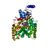
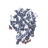
THE FIRST 6 RESIDUES (MNNRNH) AND RESIDUE 99 (Q) IN BOTH CHAINS ARE DISORDERED. IN CHAIN A RESIDUE ...THE FIRST 6 RESIDUES (MNNRNH) AND RESIDUE 99 (Q) IN BOTH CHAINS ARE DISORDERED. IN CHAIN A RESIDUE 572-577 (AEADRQ) AND IN CHAIN B REDIDUE 573-575 (EAD) ARE DISORDERED. THE TERMINAL RESIDUES 588-620 ( GHAVQHGSEVQHDERRHGDVRHEEARHGEVQHG)ARE DISORDERED IN BOTH CHAINS.

-
Experimental details

-
Experiment

ExperimentMethod: X-RAY DIFFRACTION / Number of used crystals: 1

-
Sample preparation

CrystalDensity Matthews: 2.6 Å3/Da / Density % sol: 53 % / Description: NONE
Crystal growMethod: vapor diffusion, hanging drop / pH: 6.5
Details: 29% PEG8000, 200 MM (NH4)2SO4, 10 MM SODIUM CITRATE, 5 MM ADENOSINE, 100 MM NACAC PH 6.5 ACSD CONCENTRATION: 9 MG/ML, 3UL PROTEIN AND EQUAL AMOUNTS OF PRECIPITANT USED FOR HANGING DROP METHOD

-
Data collection

DiffractionMean temperature: 100 K
Diffraction sourceSource: SYNCHROTRON / Site: ESRF / Beamline: ID23-2 / Wavelength: 0.873
DetectorType: MARRESEARCH / Detector: CCD / Date: Nov 9, 2007
Details: PT COATED MIRRORS IN A KIRKPATRICK-BAEZ (KB) GEOMETRY
RadiationMonochromator: HORIZONTALLY DIFFRACTING MONOCHROMATOR / Protocol: SINGLE WAVELENGTH / Monochromatic (M) / Laue (L): M / Scattering type: x-ray
Radiation wavelengthWavelength: 0.873 Å / Relative weight: 1
ReflectionResolution: 2.95→45.9 Å / Num. obs: 29165 / % possible obs: 98.2 % / Observed criterion σ(I): 2 / Redundancy: 2 % / Rmerge(I) obs: 0.14 / Net I/σ(I): 6.2
Reflection shellResolution: 2.95→3.11 Å / Redundancy: 2 % / Rmerge(I) obs: 0.44 / Mean I/σ(I) obs: 1.9 / % possible all: 98.5

-
Processing

Software
NameVersionClassification
REFMAC5.2.0019refinement
XDSdata reduction
SCALACCP4data scaling
PHASERphasing
RefinementMethod to determine structure: MOLECULAR REPLACEMENT
Starting model: APO STRUCTURE OF ACSD

Resolution: 2.95→91.67 Å / Cor.coef. Fo:Fc: 0.913 / Cor.coef. Fo:Fc free: 0.843 / SU B: 47.076 / SU ML: 0.405 / Cross valid method: THROUGHOUT / ESU R Free: 0.469 / Stereochemistry target values: MAXIMUM LIKELIHOOD / Details: HYDROGENS HAVE BEEN ADDED IN THE RIDING POSITIONS.
RfactorNum. reflection% reflectionSelection details
Rfree0.267 1474 5.1 %RANDOM
Rwork0.202 ---
obs0.205 27691 98.2 %-
Solvent computationIon probe radii: 0.8 Å / Shrinkage radii: 0.8 Å / VDW probe radii: 1.2 Å / Solvent model: BABINET MODEL WITH MASK
Displacement parametersBiso mean: 20.15 Å2
Baniso -1Baniso -2Baniso -3
1-1.71 Å2-3.16 Å2-3.05 Å2
2--0.22 Å20.04 Å2
3----3.68 Å2
Refinement stepCycle: LAST / Resolution: 2.95→91.67 Å
ProteinNucleic acidLigandSolventTotal
Num. atoms9227 0 61 73 9361
Refine LS restraints
Refine-IDTypeDev idealDev ideal targetNumber
X-RAY DIFFRACTIONr_bond_refined_d0.0160.0219529
X-RAY DIFFRACTIONr_bond_other_d
X-RAY DIFFRACTIONr_angle_refined_deg1.7471.95512954
X-RAY DIFFRACTIONr_angle_other_deg
X-RAY DIFFRACTIONr_dihedral_angle_1_deg7.94751145
X-RAY DIFFRACTIONr_dihedral_angle_2_deg34.30123.347484
X-RAY DIFFRACTIONr_dihedral_angle_3_deg19.351151543
X-RAY DIFFRACTIONr_dihedral_angle_4_deg19.9781587
X-RAY DIFFRACTIONr_chiral_restr0.1570.21377
X-RAY DIFFRACTIONr_gen_planes_refined0.0060.027433
X-RAY DIFFRACTIONr_gen_planes_other
X-RAY DIFFRACTIONr_nbd_refined0.2640.25210
X-RAY DIFFRACTIONr_nbd_other
X-RAY DIFFRACTIONr_nbtor_refined0.3270.26436
X-RAY DIFFRACTIONr_nbtor_other
X-RAY DIFFRACTIONr_xyhbond_nbd_refined0.1850.2469
X-RAY DIFFRACTIONr_xyhbond_nbd_other
X-RAY DIFFRACTIONr_metal_ion_refined
X-RAY DIFFRACTIONr_metal_ion_other
X-RAY DIFFRACTIONr_symmetry_vdw_refined0.2880.258
X-RAY DIFFRACTIONr_symmetry_vdw_other
X-RAY DIFFRACTIONr_symmetry_hbond_refined0.2570.22
X-RAY DIFFRACTIONr_symmetry_hbond_other
X-RAY DIFFRACTIONr_symmetry_metal_ion_refined
X-RAY DIFFRACTIONr_symmetry_metal_ion_other
X-RAY DIFFRACTIONr_mcbond_it0.5681.55853
X-RAY DIFFRACTIONr_mcbond_other
X-RAY DIFFRACTIONr_mcangle_it1.03129217
X-RAY DIFFRACTIONr_mcangle_other
X-RAY DIFFRACTIONr_scbond_it1.53934160
X-RAY DIFFRACTIONr_scbond_other
X-RAY DIFFRACTIONr_scangle_it2.5614.53737
X-RAY DIFFRACTIONr_scangle_other
X-RAY DIFFRACTIONr_long_range_B_refined
X-RAY DIFFRACTIONr_long_range_B_other
X-RAY DIFFRACTIONr_rigid_bond_restr
X-RAY DIFFRACTIONr_sphericity_free
X-RAY DIFFRACTIONr_sphericity_bonded
Refine LS restraints NCS

Refine-ID: X-RAY DIFFRACTION

Ens-IDDom-IDAuth asym-IDNumberTypeRms dev position (Å)Weight position
11A628tight positional0.070.05
12B628tight positional0.070.05
31A732tight positional0.080.05
32B732tight positional0.080.05
41A804tight positional0.080.05
42B804tight positional0.080.05
21A132medium positional0.270.5
22B132medium positional0.270.5
11A633loose positional0.65
12B633loose positional0.65
21A140loose positional0.565
22B140loose positional0.565
31A698loose positional0.545
32B698loose positional0.545
41A834loose positional0.735
42B834loose positional0.735
11A628tight thermal0.110.5
12B628tight thermal0.110.5
31A732tight thermal0.120.5
32B732tight thermal0.120.5
41A804tight thermal0.110.5
42B804tight thermal0.110.5
21A132medium thermal0.672
22B132medium thermal0.672
11A633loose thermal1.2510
12B633loose thermal1.2510
21A140loose thermal1.9810
22B140loose thermal1.9810
31A698loose thermal1.510
32B698loose thermal1.510
41A834loose thermal1.7110
42B834loose thermal1.7110
LS refinement shellResolution: 2.95→3.03 Å / Total num. of bins used: 20 /
RfactorNum. reflection
Rfree0.384 107
Rwork0.31 2055

+
About Yorodumi

-
News

-
Feb 9, 2022. New format data for meta-information of EMDB entries

New format data for meta-information of EMDB entries

  • Version 3 of the EMDB header file is now the official format.
  • The previous official version 1.9 will be removed from the archive.

Related info.:EMDB header

External links:wwPDB to switch to version 3 of the EMDB data model

-
Aug 12, 2020. Covid-19 info

Covid-19 info

URL: https://pdbj.org/emnavi/covid19.php

New page: Covid-19 featured information page in EM Navigator.

Related info.:Covid-19 info / Mar 5, 2020. Novel coronavirus structure data

+
Mar 5, 2020. Novel coronavirus structure data

Novel coronavirus structure data

Related info.:Yorodumi Speices / Aug 12, 2020. Covid-19 info

External links:COVID-19 featured content - PDBj / Molecule of the Month (242):Coronavirus Proteases

+
Jan 31, 2019. EMDB accession codes are about to change! (news from PDBe EMDB page)

EMDB accession codes are about to change! (news from PDBe EMDB page)

  • The allocation of 4 digits for EMDB accession codes will soon come to an end. Whilst these codes will remain in use, new EMDB accession codes will include an additional digit and will expand incrementally as the available range of codes is exhausted. The current 4-digit format prefixed with “EMD-” (i.e. EMD-XXXX) will advance to a 5-digit format (i.e. EMD-XXXXX), and so on. It is currently estimated that the 4-digit codes will be depleted around Spring 2019, at which point the 5-digit format will come into force.
  • The EM Navigator/Yorodumi systems omit the EMD- prefix.

Related info.:Q: What is EMD? / ID/Accession-code notation in Yorodumi/EM Navigator

External links:EMDB Accession Codes are Changing Soon! / Contact to PDBj

+
Jul 12, 2017. Major update of PDB

Major update of PDB

  • wwPDB released updated PDB data conforming to the new PDBx/mmCIF dictionary.
  • This is a major update changing the version number from 4 to 5, and with Remediation, in which all the entries are updated.
  • In this update, many items about electron microscopy experimental information are reorganized (e.g. em_software).
  • Now, EM Navigator and Yorodumi are based on the updated data.

External links:wwPDB Remediation / Enriched Model Files Conforming to OneDep Data Standards Now Available in the PDB FTP Archive

-
Yorodumi

Thousand views of thousand structures

  • Yorodumi is a browser for structure data from EMDB, PDB, SASBDB, etc.
  • This page is also the successor to EM Navigator detail page, and also detail information page/front-end page for Omokage search.
  • The word "yorodu" (or yorozu) is an old Japanese word meaning "ten thousand". "mi" (miru) is to see.

Related info.:EMDB / PDB / SASBDB / Comparison of 3 databanks / Yorodumi Search / Aug 31, 2016. New EM Navigator & Yorodumi / Yorodumi Papers / Jmol/JSmol / Function and homology information / Changes in new EM Navigator and Yorodumi

Read more