primosome complex / DNA replication, synthesis of primer / DNA helicase activity / DNA helicase / ATP hydrolysis activity / DNA binding / ATP binding Similarity search - Function
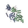
DNAb Helicase; Chain A / DNAb Helicase; Chain A / DNA helicase, DnaB type / DNA helicase, DnaB-like, N-terminal / DnaB-like helicase N terminal domain / DNA helicase, DnaB-like, N-terminal domain superfamily / DNA helicase DnaB, N-terminal/DNA primase DnaG, C-terminal / DnaB-like helicase C terminal domain / DNA helicase, DnaB-like, C-terminal / Superfamily 4 helicase domain profile. ...DNAb Helicase; Chain A / DNAb Helicase; Chain A / DNA helicase, DnaB type / DNA helicase, DnaB-like, N-terminal / DnaB-like helicase N terminal domain / DNA helicase, DnaB-like, N-terminal domain superfamily / DNA helicase DnaB, N-terminal/DNA primase DnaG, C-terminal / DnaB-like helicase C terminal domain / DNA helicase, DnaB-like, C-terminal / Superfamily 4 helicase domain profile. / P-loop containing nucleotide triphosphate hydrolases / P-loop containing nucleoside triphosphate hydrolase / Rossmann fold / Orthogonal Bundle / 3-Layer(aba) Sandwich / Mainly Alpha / Alpha Beta Similarity search - Domain/homology
Resolution: 3.6→30 Å / Num. obs: 18502 / % possible obs: 99.2 % / Observed criterion σ(I): 2 / Redundancy: 5.1 % / Rmerge(I) obs: 0.06 / Net I/σ(I): 36.4
Reflection shell
Resolution: 3.6→3.73 Å / Redundancy: 4.2 % / Rmerge(I) obs: 0.68 / Mean I/σ(I) obs: 2.2 / % possible all: 95.5
-
Processing
Software
Name
Version
Classification
CNS
1.2
refinement
SCALEPACK
datascaling
SOLVE
phasing
Refinement
Method to determine structure: MAD Starting model: NONE Resolution: 3.6→24.87 Å / Rfactor Rfree error: 0.009 / Isotropic thermal model: RESTRAINED / Cross valid method: THROUGHOUT / σ(F): 2 Details: THE RESIDUES IN CHAIN A INCLUDING 1- 7, 258-264, 365-381, 400-409,AND 436-454 WERE MISSING. THE RESIDUES IN CHAIN B INCLUDING 1-6, 155-157, 179-182, 322-331, 365-381, 397-410 AND 426-454 WERE MISSING.
In the structure databanks used in Yorodumi, some data are registered as the other names, "COVID-19 virus" and "2019-nCoV". Here are the details of the virus and the list of structure data.
Jan 31, 2019. EMDB accession codes are about to change! (news from PDBe EMDB page)
EMDB accession codes are about to change! (news from PDBe EMDB page)
The allocation of 4 digits for EMDB accession codes will soon come to an end. Whilst these codes will remain in use, new EMDB accession codes will include an additional digit and will expand incrementally as the available range of codes is exhausted. The current 4-digit format prefixed with “EMD-” (i.e. EMD-XXXX) will advance to a 5-digit format (i.e. EMD-XXXXX), and so on. It is currently estimated that the 4-digit codes will be depleted around Spring 2019, at which point the 5-digit format will come into force.
The EM Navigator/Yorodumi systems omit the EMD- prefix.
Related info.:Q: What is EMD? / ID/Accession-code notation in Yorodumi/EM Navigator
Yorodumi is a browser for structure data from EMDB, PDB, SASBDB, etc.
This page is also the successor to EM Navigator detail page, and also detail information page/front-end page for Omokage search.
The word "yorodu" (or yorozu) is an old Japanese word meaning "ten thousand". "mi" (miru) is to see.
Related info.:EMDB / PDB / SASBDB / Comparison of 3 databanks / Yorodumi Search / Aug 31, 2016. New EM Navigator & Yorodumi / Yorodumi Papers / Jmol/JSmol / Function and homology information / Changes in new EM Navigator and Yorodumi
 Movie
Movie Controller


 Open data
 Basic information
 Components
 Keywords
 HYDROLASE /
  Function and homology information
 GEOBACILLUS KAUSTOPHILUS HTA426 (bacteria)
 Authors
 Citation
 Structure visualization
 Downloads & links
 Other downloads


 PDBj
PDBj


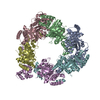
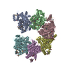
 Assembly
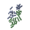
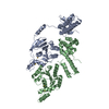

 Mass: 196.967 Da / Num. of mol.: 4 / Source method: obtained synthetically / Formula: Au
 Sample preparation
 / Beamline: BL13B1 / Wavelength: 1
  Processing