 Movie
Movie Controller


+ Open data

- Basic information
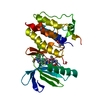
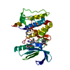
| Entry | Database: PDB / ID: 2vwu | ||||||
|---|---|---|---|---|---|---|---|
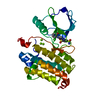
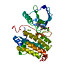
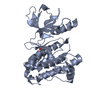
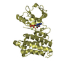
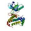
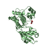
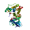
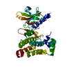
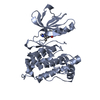
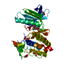
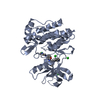
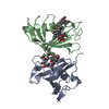
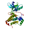
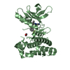
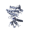
| Title | ephB4 kinase domain inhibitor complex | ||||||
 Components Components | EPHRIN TYPE-B RECEPTOR 4 | ||||||
 Keywords Keywords |  TRANSFERASE / TYROSINE-PROTEIN KINASE / RECEPTOR TYROSINE KINASE / KINASE / MUTANT / MEMBRANE / RECEPTOR / ATP-BINDING / POLYMORPHISM / GLYCOPROTEIN / TRANSMEMBRANE / PHOSPHOPROTEIN / UNPHOSPHORYLATED / NUCLEOTIDE-BINDING TRANSFERASE / TYROSINE-PROTEIN KINASE / RECEPTOR TYROSINE KINASE / KINASE / MUTANT / MEMBRANE / RECEPTOR / ATP-BINDING / POLYMORPHISM / GLYCOPROTEIN / TRANSMEMBRANE / PHOSPHOPROTEIN / UNPHOSPHORYLATED / NUCLEOTIDE-BINDING | ||||||
| Function / homology |  Function and homology informationephrin receptor activity / cell migration involved in sprouting angiogenesis / EPH-Ephrin signaling / Ephrin signaling / EPH-ephrin mediated repulsion of cells / ephrin receptor signaling pathway / heart morphogenesis / EPHB-mediated forward signaling / transmembrane receptor protein tyrosine kinase activity / receptor protein-tyrosine kinase ...ephrin receptor activity / cell migration involved in sprouting angiogenesis / EPH-Ephrin signaling / Ephrin signaling / EPH-ephrin mediated repulsion of cells / ephrin receptor signaling pathway / heart morphogenesis / EPHB-mediated forward signaling / transmembrane receptor protein tyrosine kinase activity / receptor protein-tyrosine kinase / angiogenesis / protein autophosphorylation / receptor complex / cell adhesion / extracellular exosome / extracellular region / ATP binding / plasma membrane / cytosol Function and homology informationephrin receptor activity / cell migration involved in sprouting angiogenesis / EPH-Ephrin signaling / Ephrin signaling / EPH-ephrin mediated repulsion of cells / ephrin receptor signaling pathway / heart morphogenesis / EPHB-mediated forward signaling / transmembrane receptor protein tyrosine kinase activity / receptor protein-tyrosine kinase ...ephrin receptor activity / cell migration involved in sprouting angiogenesis / EPH-Ephrin signaling / Ephrin signaling / EPH-ephrin mediated repulsion of cells / ephrin receptor signaling pathway / heart morphogenesis / EPHB-mediated forward signaling / transmembrane receptor protein tyrosine kinase activity / receptor protein-tyrosine kinase / angiogenesis / protein autophosphorylation / receptor complex / cell adhesion / extracellular exosome / extracellular region / ATP binding / plasma membrane / cytosolSimilarity search - Function | ||||||
| Biological species |  HOMO SAPIENS (human) HOMO SAPIENS (human) | ||||||
| Method | X-RAY DIFFRACTION / SYNCHROTRON / MOLECULAR REPLACEMENT / Resolution: 2 Å | ||||||
 Authors Authors | Read, J. / Brassington, C.A. / Green, I. / McCall, E.J. / Valentine, A.L. / Barratt, D. / Rowsell, S. / Packer, M. / McAlister, M. | ||||||
 Citation Citation | Journal: Bioorg.Med.Chem.Lett. / Year: 2008 Title: Inhibitors of the Tyrosine Kinase Ephb4. Part 1: Structure-Based Design and Optimization of a Series of 2,4-Bis-Anilinopyrimidines Authors: Bardelle, C. / Cross, D. / Davenport, S. / Kettle, J.G. / Ko, E.J. / Leach, A.G. / Mortlock, A. / Read, J. / Roberts, N.J. / Robins, P. / Williams, E.J. | ||||||
| History |
|
- Structure visualization
| Structure viewer | Molecule: MolmilJmol/JSmol |
|---|
- Downloads & links
-Download
| PDBx/mmCIF format | 2vwu.cif.gz | 68.7 KB | Display | PDBx/mmCIF format |
|---|---|---|---|---|
| PDB format | pdb2vwu.ent.gz | 48.2 KB | Display | PDB format |
| PDBx/mmJSON format | 2vwu.json.gz | Tree view | PDBx/mmJSON format | |
| Others |  Other downloads Other downloads |
-Validation report
| Arichive directory | https://data.pdbj.org/pub/pdb/validation_reports/vw/2vwuftp://data.pdbj.org/pub/pdb/validation_reports/vw/2vwu | HTTPS FTP |
|---|
-Related structure data
| Related structure data |  2vwvC  2vwwC 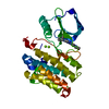 2vx0C  1jpaS C: citing same article ( S: Starting model for refinement |
|---|---|
| Similar structure data |


-Links
 PDBj
PDBj


- Assembly
| Deposited unit | 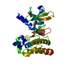
| ||||||||
|---|---|---|---|---|---|---|---|---|---|
| 1 |
| ||||||||
| Unit cell |
|
-Components
| #1: Protein | Mass: 33934.816 Da / Num. of mol.: 1 / Fragment: KINASE DOMAIN, RESIDUES 598-887 / Mutation: YES Source method: isolated from a genetically manipulated source Source: (gene. exp.) HOMO SAPIENS (human) / Plasmid: PFASTBAC / Cell line (production host): Sf21 / Production host:   SPODOPTERA FRUGIPERDA (fall armyworm) SPODOPTERA FRUGIPERDA (fall armyworm)References: UniProt: P54760, receptor protein-tyrosine kinase | ||
|---|---|---|---|
| #2: Chemical | ChemComp-7X1 /   Mass: 470.949 Da / Num. of mol.: 1 / Source method: obtained synthetically / Formula: C24H27ClN4O4 Mass: 470.949 Da / Num. of mol.: 1 / Source method: obtained synthetically / Formula: C24H27ClN4O4 | ||
| #3: Water | ChemComp-HOH / Water Mass: 18.015 Da / Num. of mol.: 127 / Source method: isolated from a natural source / Formula: H2O Mass: 18.015 Da / Num. of mol.: 127 / Source method: isolated from a natural source / Formula: H2O | ||
| Compound details | ENGINEERED| Sequence details | Y774E MUTATION NOT SEEN IN ELECTRON DENSITY | |
-Experimental details
-Experiment
| Experiment | Method: X-RAY DIFFRACTION / Number of used crystals: 1 |
|---|
- Sample preparation
| Crystal | Density Matthews: 2.4 Å3/Da / Density % sol: 48.36 % / Description: NONE |
|---|---|
| Crystal grow | Temperature: 277 K / Method: vapor diffusion, sitting drop / pH: 7.5 Details: PROTEIN: 12MG/ML IN 50MM MOPS PH 6.5, 50MM NACL, 1MM DTT RESERVOIR: 25% PEG 5000 MME, 0.1M TRIS PH 7.5, 0.15M MGCL2, 15% GLYCEROL TEMP: 4 DEGREES C SITTING DROP: 2 UL PROTEIN, 0.6 UL RESERVOIR |
-Data collection
| Diffraction | Mean temperature: 100 K |
|---|---|
| Diffraction source | Source: SYNCHROTRON / Site: ESRF  / Beamline: ID29 / Wavelength: 0.97568 / Beamline: ID29 / Wavelength: 0.97568 |
| Detector | Type: ADSC CCD / Detector: CCD / Date: Nov 24, 2004 / Details: MIRRORS |
| Radiation | Protocol: SINGLE WAVELENGTH / Monochromatic (M) / Laue (L): M / Scattering type: x-ray |
| Radiation wavelength | Wavelength: 0.97568 Å / Relative weight: 1 |
| Reflection | Resolution: 1.45→56.8 Å / Num. obs: 19173 / % possible obs: 100 % / Observed criterion σ(I): 2 / Redundancy: 7.1 % / Biso Wilson estimate: 16.18 Å2 / Rmerge(I) obs: 0.09 / Net I/σ(I): 5.44 |
| Reflection shell | Resolution: 2→2.11 Å / Redundancy: 7.15 % / Rmerge(I) obs: 0.38 / Mean I/σ(I) obs: 1.6 / % possible all: 100 |
- Processing
| Software |
| ||||||||||||||||||||||||||||||||||||||||||||||||||||||||||||||||||||||||||||||||||||||||||||||||||||||||||||||||||||||||||||||||||||||||||||||||||||||||||||||||||||||||||||||||||||||
|---|---|---|---|---|---|---|---|---|---|---|---|---|---|---|---|---|---|---|---|---|---|---|---|---|---|---|---|---|---|---|---|---|---|---|---|---|---|---|---|---|---|---|---|---|---|---|---|---|---|---|---|---|---|---|---|---|---|---|---|---|---|---|---|---|---|---|---|---|---|---|---|---|---|---|---|---|---|---|---|---|---|---|---|---|---|---|---|---|---|---|---|---|---|---|---|---|---|---|---|---|---|---|---|---|---|---|---|---|---|---|---|---|---|---|---|---|---|---|---|---|---|---|---|---|---|---|---|---|---|---|---|---|---|---|---|---|---|---|---|---|---|---|---|---|---|---|---|---|---|---|---|---|---|---|---|---|---|---|---|---|---|---|---|---|---|---|---|---|---|---|---|---|---|---|---|---|---|---|---|---|---|---|---|
| Refinement | Method to determine structure: MOLECULAR REPLACEMENT Starting model: PDB ENTRY 1JPA Resolution: 2→56.8 Å / Cor.coef. Fo:Fc: 0.946 / Cor.coef. Fo:Fc free: 0.935 / SU B: 7.218 / SU ML: 0.094 / Cross valid method: THROUGHOUT / σ(F): 2 / ESU R: 0.17 / ESU R Free: 0.147 / Stereochemistry target values: MAXIMUM LIKELIHOOD Details: HYDROGENS HAVE BEEN ADDED IN THE RIDING POSITIONS. THE FOLLOWING RESIDUES ARE DISORDERED 598 TO 608, 649 TO 653, 760 TO 781 AND 888 TO 892
| ||||||||||||||||||||||||||||||||||||||||||||||||||||||||||||||||||||||||||||||||||||||||||||||||||||||||||||||||||||||||||||||||||||||||||||||||||||||||||||||||||||||||||||||||||||||
| Solvent computation | Ion probe radii: 0.8 Å / VDW probe radii: 1.2 Å / Solvent model: BABINET MODEL PLUS MASK | ||||||||||||||||||||||||||||||||||||||||||||||||||||||||||||||||||||||||||||||||||||||||||||||||||||||||||||||||||||||||||||||||||||||||||||||||||||||||||||||||||||||||||||||||||||||
| Displacement parameters | Biso mean: 23.29 Å2
| ||||||||||||||||||||||||||||||||||||||||||||||||||||||||||||||||||||||||||||||||||||||||||||||||||||||||||||||||||||||||||||||||||||||||||||||||||||||||||||||||||||||||||||||||||||||
| Refinement step | Cycle: LAST / Resolution: 2→56.8 Å
| ||||||||||||||||||||||||||||||||||||||||||||||||||||||||||||||||||||||||||||||||||||||||||||||||||||||||||||||||||||||||||||||||||||||||||||||||||||||||||||||||||||||||||||||||||||||
| Refine LS restraints |
|