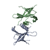
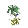
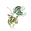
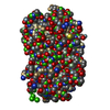
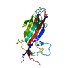
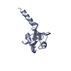
Entry Database : PDB / ID : 2vsvTitle Crystal structure of the PDZ domain of human rhophilin-2 RHOPHILIN-2 Keywords / / / / / / / / Function / homology Function Domain/homology Component
/ / / / / / / / / / / / / / / / / / / / / / / / / Biological species HOMO SAPIENS (human)Method / / / Resolution : 1.82 Å Authors Pike, A.C.W. / Kochan, G. / Sun, Z. / Shafqat, N. / Pilka, E.S. / Roos, A. / Elkins, J. / Burgess-Brown, N. / Murray, J.W. / von Delft, F. ...Pike, A.C.W. / Kochan, G. / Sun, Z. / Shafqat, N. / Pilka, E.S. / Roos, A. / Elkins, J. / Burgess-Brown, N. / Murray, J.W. / von Delft, F. / Wikstrom, M. / Edwards, A. / Arrowsmith, C.H. / Bountra, C. / Oppermann, U. Journal : To be Published Title : Crystal Structure of the Pdz Domain of Human Rhophilin-2Authors: Pike, A.C.W. / Kochan, G. / Sun, Z. / Shafqat, N. / Pilka, E.S. / Roos, A. / Elkins, J. / Burgess-Brown, N. / Murray, J.W. / von Delft, F. / Wikstrom, M. / Edwards, A. / Arrowsmith, C.H. / ... Authors : Pike, A.C.W. / Kochan, G. / Sun, Z. / Shafqat, N. / Pilka, E.S. / Roos, A. / Elkins, J. / Burgess-Brown, N. / Murray, J.W. / von Delft, F. / Wikstrom, M. / Edwards, A. / Arrowsmith, C.H. / Bountra, C. / Oppermann, U. History Deposition Apr 29, 2008 Deposition site / Processing site Revision 1.0 Jul 15, 2008 Provider / Type Revision 1.1 May 8, 2011 Group Revision 1.2 Jul 13, 2011 Group Revision 1.3 Jan 24, 2018 Group / Category / Item Revision 1.4 Dec 13, 2023 Group Data collection / Database references ... Data collection / Database references / Other / Refinement description Category chem_comp_atom / chem_comp_bond ... chem_comp_atom / chem_comp_bond / database_2 / pdbx_database_status / pdbx_initial_refinement_model Item / _database_2.pdbx_database_accession / _pdbx_database_status.status_code_sf
Show all Show less
 Movie
Movie Controller


 Open data
 Basic information
 Components
 Keywords
 PROTEIN BINDING /
  Function and homology information
 Authors
 Citation
 Structure visualization
 Downloads & links
 Other downloads
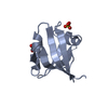


 PDBj
PDBj Assembly
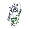


 Mass: 18.015 Da / Num. of mol.: 93 / Source method: isolated from a natural source / Formula: H2O
 Sample preparation
 / Beamline: X10SA / Wavelength: 0.99186
  Processing