 Movie
Movie Controller

[English] 日本語
 Yorodumi
- PDB-2vqo: Structure of HDAC4 catalytic domain with a gain-of-function muati... -
+ Open data

- Basic information
| Entry | Database: PDB / ID: 2vqo | ||||||
|---|---|---|---|---|---|---|---|
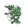
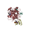
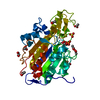
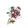
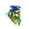
| Title | Structure of HDAC4 catalytic domain with a gain-of-function muation bound to a trifluoromethylketone inhbitor | ||||||
 Components Components | HISTONE DEACETYLASE 4 | ||||||
 Keywords Keywords | HYDROLASE / INHIBITOR / REPRESSOR / CHROMATIN / COILED COIL / HISTONE DEACETYLASE / TRANSCRIPTION REGULATION / UBL CONJUGATION / CHROMATIN REGULATOR / POLYMORPHISM / TRANSCRIPTION / PHOSPHOPROTEIN / HDAC / ZINC / HDACI / NUCLEUS / CYTOPLASM | ||||||
| Function / homology |  Function and homology information Function and homology informationRUNX2 regulates chondrocyte maturation / response to denervation involved in regulation of muscle adaptation / negative regulation of myotube differentiation / peptidyl-lysine deacetylation / positive regulation of protein sumoylation / negative regulation of transcription by competitive promoter binding / regulation of protein binding / protein deacetylation / cardiac muscle hypertrophy in response to stress / histone deacetylase ...RUNX2 regulates chondrocyte maturation / response to denervation involved in regulation of muscle adaptation / negative regulation of myotube differentiation / peptidyl-lysine deacetylation / positive regulation of protein sumoylation / negative regulation of transcription by competitive promoter binding / regulation of protein binding / protein deacetylation / cardiac muscle hypertrophy in response to stress / histone deacetylase / protein lysine deacetylase activity / negative regulation of glycolytic process / SUMO transferase activity / histone deacetylase activity / negative regulation of gene expression, epigenetic / B cell activation / type I interferon-mediated signaling pathway / Notch-HLH transcription pathway / potassium ion binding / protein sumoylation / histone deacetylase complex / RUNX3 regulates p14-ARF / transcription repressor complex / SUMOylation of chromatin organization proteins / response to interleukin-1 / B cell differentiation / SUMOylation of intracellular receptors / negative regulation of DNA-binding transcription factor activity / NOTCH1 Intracellular Domain Regulates Transcription / Constitutive Signaling by NOTCH1 PEST Domain Mutants / Constitutive Signaling by NOTCH1 HD+PEST Domain Mutants / histone deacetylase binding / positive regulation of DNA-binding transcription factor activity / nervous system development / DNA-binding transcription factor binding / RNA polymerase II-specific DNA-binding transcription factor binding / molecular adaptor activity / nuclear speck / chromatin remodeling / inflammatory response / RNA polymerase II cis-regulatory region sequence-specific DNA binding / positive regulation of cell population proliferation / chromatin / positive regulation of DNA-templated transcription / negative regulation of transcription by RNA polymerase II / positive regulation of transcription by RNA polymerase II / zinc ion binding / nucleoplasm / identical protein binding / nucleus / cytosol / cytoplasmSimilarity search - Function | ||||||
| Biological species |  HOMO SAPIENS (human) HOMO SAPIENS (human) | ||||||
| Method | X-RAY DIFFRACTION / SYNCHROTRON / MOLECULAR REPLACEMENT / Resolution: 2.15 Å | ||||||
 Authors Authors | Bottomley, M.J. / Lo Surdo, P. / Di Giovine, P. / Cirillo, A. / Scarpelli, R. / Ferrigno, F. / Jones, P. / Neddermann, P. / De Francesco, R. / Steinkuhler, C. ...Bottomley, M.J. / Lo Surdo, P. / Di Giovine, P. / Cirillo, A. / Scarpelli, R. / Ferrigno, F. / Jones, P. / Neddermann, P. / De Francesco, R. / Steinkuhler, C. / Gallinari, P. / Carfi, A. | ||||||
 Citation Citation | Journal: J.Biol.Chem. / Year: 2008 Title: Structural and Functional Analysis of the Human Hdac4 Catalytic Domain Reveals a Regulatory Zinc-Binding Domain. Authors: Bottomley, M.J. / Lo Surdo, P. / Di Giovine, P. / Cirillo, A. / Scarpelli, R. / Ferrigno, F. / Jones, P. / Neddermann, P. / De Francesco, R. / Steinkuhler, C. / Gallinari, P. / Carfi, A. | ||||||
| History |
|
- Structure visualization
| Structure viewer | Molecule: MolmilJmol/JSmol |
|---|
- Downloads & links
-Download
| PDBx/mmCIF format | 2vqo.cif.gz | 156.1 KB | Display | PDBx/mmCIF format |
|---|---|---|---|---|
| PDB format | pdb2vqo.ent.gz | 121.5 KB | Display | PDB format |
| PDBx/mmJSON format | 2vqo.json.gz | Tree view | PDBx/mmJSON format | |
| Others |  Other downloads Other downloads |
-Validation report
| Arichive directory | https://data.pdbj.org/pub/pdb/validation_reports/vq/2vqoftp://data.pdbj.org/pub/pdb/validation_reports/vq/2vqo | HTTPS FTP |
|---|
-Related structure data
| Related structure data |  2vqjSC  2vqmC  2vqqC  2vqvC  2vqwC C: citing same article ( S: Starting model for refinement |
|---|---|
| Similar structure data |


-Links
 PDBj
PDBj


- Assembly
| Deposited unit | 
| ||||||||
|---|---|---|---|---|---|---|---|---|---|
| 1 | 
| ||||||||
| 2 | 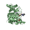
| ||||||||
| Unit cell |
|
-Components
-Protein , 1 types, 2 molecules AB
| #1: Protein | / HD4 Mass: 44438.301 Da / Num. of mol.: 2 / Fragment: CATALYTIC DOMAIN, RESIDUES 648-1057 / Mutation: YES Source method: isolated from a genetically manipulated source Source: (gene. exp.) HOMO SAPIENS (human) / Plasmid: PETM-11 (OBTAINED FROM EMBL-HEIDELBERG) / Production host:  ESCHERICHIA COLI (E. coli) / Strain (production host): BL21(DE3) / References: UniProt: P56524 ESCHERICHIA COLI (E. coli) / Strain (production host): BL21(DE3) / References: UniProt: P56524 |
|---|
-Non-polymers , 5 types, 263 molecules 


| #2: Chemical | Sulfate Mass: 96.063 Da / Num. of mol.: 3 / Source method: obtained synthetically / Formula: SO4 Mass: 96.063 Da / Num. of mol.: 3 / Source method: obtained synthetically / Formula: SO4#3: Chemical | ChemComp-K /  Mass: 39.098 Da / Num. of mol.: 4 / Source method: obtained synthetically / Formula: K Mass: 39.098 Da / Num. of mol.: 4 / Source method: obtained synthetically / Formula: K#4: Chemical |  Mass: 423.409 Da / Num. of mol.: 2 / Source method: obtained synthetically / Formula: C19H16F3N3O3S Mass: 423.409 Da / Num. of mol.: 2 / Source method: obtained synthetically / Formula: C19H16F3N3O3S#5: Chemical |  Mass: 65.409 Da / Num. of mol.: 2 / Source method: obtained synthetically / Formula: Zn Mass: 65.409 Da / Num. of mol.: 2 / Source method: obtained synthetically / Formula: Zn#6: Water | ChemComp-HOH / | WaterMass: 18.015 Da / Num. of mol.: 252 / Source method: isolated from a natural source / Formula: H2O |
|---|
-Details
| Compound details | ENGINEERED RESIDUE IN CHAIN A, CYS 669 TO ALA ENGINEERED RESIDUE IN CHAIN A, CYS 700 TO ALA ...ENGINEERED |
|---|
-Experimental details
-Experiment
| Experiment | Method: X-RAY DIFFRACTION / Number of used crystals: 1 |
|---|
- Sample preparation
| Crystal | Density Matthews: 2.9 Å3/Da / Density % sol: 57 % / Description: NONE |
|---|---|
| Crystal grow | pH: 6.5 Details: 1.6M AMMONIUM SULPHATE, 0.1M MES PH 6.5, 10% DIOXANE, 1MM DTT |
-Data collection
| Diffraction | Mean temperature: 100 K |
|---|---|
| Diffraction source | Source: SYNCHROTRON / Site: ESRF  / Beamline: ID14-1 / Wavelength: 0.94 / Beamline: ID14-1 / Wavelength: 0.94 |
| Detector | Type: ADSC CCD / Detector: CCD / Date: Jan 12, 2007 |
| Radiation | Protocol: SINGLE WAVELENGTH / Monochromatic (M) / Laue (L): M / Scattering type: x-ray |
| Radiation wavelength | Wavelength: 0.94 Å / Relative weight: 1 |
| Reflection | Resolution: 2.15→40 Å / Num. obs: 15329 / % possible obs: 97.5 % / Redundancy: 3 % / Rmerge(I) obs: 0.13 / Net I/σ(I): 7.8 |
| Reflection shell | Resolution: 2.15→2.21 Å / Redundancy: 3 % / Rmerge(I) obs: 0.58 / Mean I/σ(I) obs: 1.5 / % possible all: 97.5 |
- Processing
| Software |
| ||||||||||||||||||||||||||||||||||||||||||||||||||||||||||||||||||||||||||||||||||||||||||||||||||||||||||||||||||||||||||||||||||||||||||||||||||||||||||||||||||||||||||||||||||||||
|---|---|---|---|---|---|---|---|---|---|---|---|---|---|---|---|---|---|---|---|---|---|---|---|---|---|---|---|---|---|---|---|---|---|---|---|---|---|---|---|---|---|---|---|---|---|---|---|---|---|---|---|---|---|---|---|---|---|---|---|---|---|---|---|---|---|---|---|---|---|---|---|---|---|---|---|---|---|---|---|---|---|---|---|---|---|---|---|---|---|---|---|---|---|---|---|---|---|---|---|---|---|---|---|---|---|---|---|---|---|---|---|---|---|---|---|---|---|---|---|---|---|---|---|---|---|---|---|---|---|---|---|---|---|---|---|---|---|---|---|---|---|---|---|---|---|---|---|---|---|---|---|---|---|---|---|---|---|---|---|---|---|---|---|---|---|---|---|---|---|---|---|---|---|---|---|---|---|---|---|---|---|---|---|
| Refinement | Method to determine structure: MOLECULAR REPLACEMENT Starting model: PDB ENTRY 2VQJ Resolution: 2.15→40 Å / Cor.coef. Fo:Fc: 0.947 / Cor.coef. Fo:Fc free: 0.922 / SU B: 6.024 / SU ML: 0.152 / Cross valid method: THROUGHOUT / ESU R: 0.212 / ESU R Free: 0.186 / Stereochemistry target values: MAXIMUM LIKELIHOOD Details: HYDROGENS HAVE BEEN ADDED IN THE RIDING POSITIONS. RESIDUES 23-34 AND 85-111 ARE NOT VISIBLE IN THE ELECTRON DENSITY MAPS.
| ||||||||||||||||||||||||||||||||||||||||||||||||||||||||||||||||||||||||||||||||||||||||||||||||||||||||||||||||||||||||||||||||||||||||||||||||||||||||||||||||||||||||||||||||||||||
| Solvent computation | Ion probe radii: 0.8 Å / Shrinkage radii: 0.8 Å / VDW probe radii: 1.4 Å / Solvent model: MASK | ||||||||||||||||||||||||||||||||||||||||||||||||||||||||||||||||||||||||||||||||||||||||||||||||||||||||||||||||||||||||||||||||||||||||||||||||||||||||||||||||||||||||||||||||||||||
| Displacement parameters | Biso mean: 28.02 Å2
| ||||||||||||||||||||||||||||||||||||||||||||||||||||||||||||||||||||||||||||||||||||||||||||||||||||||||||||||||||||||||||||||||||||||||||||||||||||||||||||||||||||||||||||||||||||||
| Refinement step | Cycle: LAST / Resolution: 2.15→40 Å
| ||||||||||||||||||||||||||||||||||||||||||||||||||||||||||||||||||||||||||||||||||||||||||||||||||||||||||||||||||||||||||||||||||||||||||||||||||||||||||||||||||||||||||||||||||||||
| Refine LS restraints |
|