 Movie
Movie Controller

[English] 日本語
 Yorodumi
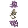
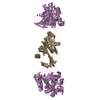
- PDB-2sic: REFINED CRYSTAL STRUCTURE OF THE COMPLEX OF SUBTILISIN BPN' AND S... -
+ Open data

- Basic information
| Entry | Database: PDB / ID: 2sic | |||||||||
|---|---|---|---|---|---|---|---|---|---|---|
| Title | REFINED CRYSTAL STRUCTURE OF THE COMPLEX OF SUBTILISIN BPN' AND STREPTOMYCES SUBTILISIN INHIBITOR AT 1.8 ANGSTROMS RESOLUTION | |||||||||
 Components Components |
| |||||||||
 Keywords Keywords | COMPLEX (PROTEINASE/INHIBITOR) / COMPLEX (PROTEINASE-INHIBITOR) / COMPLEX (PROTEINASE-INHIBITOR) complex | |||||||||
| Function / homology |  Function and homology information Function and homology information subtilisin / sporulation resulting in formation of a cellular spore / fibrinolysis / serine-type endopeptidase inhibitor activity / serine-type endopeptidase activity / proteolysis / extracellular region / metal ion binding subtilisin / sporulation resulting in formation of a cellular spore / fibrinolysis / serine-type endopeptidase inhibitor activity / serine-type endopeptidase activity / proteolysis / extracellular region / metal ion bindingSimilarity search - Function | |||||||||
| Biological species |  Bacillus amyloliquefaciens (bacteria) Bacillus amyloliquefaciens (bacteria) | |||||||||
| Method | X-RAY DIFFRACTION / Resolution: 1.8 Å | |||||||||
 Authors Authors | Mitsui, Y. / Takeuchi, Y. / Hirono, S. / Akagawa, H. / Nakamura, K.T. | |||||||||
 Citation Citation | Journal: J.Mol.Biol. / Year: 1991 Title: Refined crystal structure of the complex of subtilisin BPN' and Streptomyces subtilisin inhibitor at 1.8 A resolution. Authors: Takeuchi, Y. / Satow, Y. / Nakamura, K.T. / Mitsui, Y. #1: Journal: J.Mol.Biol. / Year: 1984Title: Crystal Structure at 2.6 Angstroms Resolution of the Complex of Subtilisin with Bpn' with Streptomyces Subtilisin Inhibitor Authors: Hirono, S. / Akagawa, H. / Mitsui, Y. / Iitaka, Y. #2: Journal: J.Mol.Biol. / Year: 1979Title: Crystal Structure of the Complex of Subtilisin Bpn' with its Protein Inhibitor Streptomyces Subtilisin Inhibitor Authors: Hirono, S. / Nakamura, K.T. / Iitaka, Y. / Mitsui, Y. #3: Journal: Nature / Year: 1979Title: Crystal Structures of Streptomyces Subtilisin Inhibitor and its Complex with Subtilisin Bpn' Authors: Mitsui, Y. / Satow, Y. / Watanabe, Y. / Hirono, S. / Iitaka, Y. | |||||||||
| History |
|
- Structure visualization
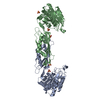
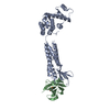
| Structure viewer | Molecule: MolmilJmol/JSmol |
|---|
- Downloads & links
-Download
| PDBx/mmCIF format | 2sic.cif.gz | 83.6 KB | Display | PDBx/mmCIF format |
|---|---|---|---|---|
| PDB format | pdb2sic.ent.gz | 65.6 KB | Display | PDB format |
| PDBx/mmJSON format | 2sic.json.gz | Tree view | PDBx/mmJSON format | |
| Others |  Other downloads Other downloads |
-Validation report
| Arichive directory | https://data.pdbj.org/pub/pdb/validation_reports/si/2sicftp://data.pdbj.org/pub/pdb/validation_reports/si/2sic | HTTPS FTP |
|---|
-Related structure data
| Similar structure data |
|---|


-Links
 PDBj
PDBj

- Assembly
| Deposited unit | 
| ||||||||
|---|---|---|---|---|---|---|---|---|---|
| 1 | 
| ||||||||
| Unit cell |
| ||||||||
| Atom site foot note | 1: RESIDUES PRO E 168 AND PRO I 37 ARE CIS PROLINES. |
-Components
| #1: Protein | Mass: 27552.525 Da / Num. of mol.: 1 Source method: isolated from a genetically manipulated source Source: (gene. exp.) Bacillus amyloliquefaciens (bacteria) / References: UniProt: P00782, 3.4.21.14 | ||
|---|---|---|---|
| #2: Protein | Mass: 10940.334 Da / Num. of mol.: 1 Source method: isolated from a genetically manipulated source References: UniProt: P01006 | ||
| #3: Chemical |   Mass: 40.078 Da / Num. of mol.: 2 / Source method: obtained synthetically / Formula: Ca Mass: 40.078 Da / Num. of mol.: 2 / Source method: obtained synthetically / Formula: Ca#4: Water | ChemComp-HOH / | Water Mass: 18.015 Da / Num. of mol.: 258 / Source method: isolated from a natural source / Formula: H2O Mass: 18.015 Da / Num. of mol.: 258 / Source method: isolated from a natural source / Formula: H2O |
-Experimental details
-Experiment
| Experiment | Method: X-RAY DIFFRACTION |
|---|
- Sample preparation
| Crystal | Density Matthews: 3.24 Å3/Da / Density % sol: 62.02 % | ||||||||||||||||||||||||
|---|---|---|---|---|---|---|---|---|---|---|---|---|---|---|---|---|---|---|---|---|---|---|---|---|---|
| Crystal grow | *PLUS pH: 7.5 / Method: unknownDetails: used seeding, Hirono, S., (1979) J.Mol.Biol., 131, 855. | ||||||||||||||||||||||||
| Components of the solutions | *PLUS
|
-Data collection
| Radiation | Scattering type: x-ray |
|---|---|
| Radiation wavelength | Relative weight: 1 |
| Reflection | *PLUS Highest resolution: 1.8 Å / Num. obs: 28118 / Observed criterion σ(F): 2 / Num. measured all: 97766 / Rmerge(I) obs: 0.0472 |
- Processing
| Software | Name: PROLSQ / Classification: refinement | ||||||||||||||||||||||||||||||||||||||||||||||||||||||||||||||||||||||||||||||||||||
|---|---|---|---|---|---|---|---|---|---|---|---|---|---|---|---|---|---|---|---|---|---|---|---|---|---|---|---|---|---|---|---|---|---|---|---|---|---|---|---|---|---|---|---|---|---|---|---|---|---|---|---|---|---|---|---|---|---|---|---|---|---|---|---|---|---|---|---|---|---|---|---|---|---|---|---|---|---|---|---|---|---|---|---|---|---|
| Refinement | Resolution: 1.8→6 Å /
| ||||||||||||||||||||||||||||||||||||||||||||||||||||||||||||||||||||||||||||||||||||
| Refinement step | Cycle: LAST / Resolution: 1.8→6 Å
| ||||||||||||||||||||||||||||||||||||||||||||||||||||||||||||||||||||||||||||||||||||
| Refine LS restraints |
|
