 Movie
Movie Controller


+ Open data

- Basic information
| Entry | Database: PDB / ID: 2rdo | ||||||
|---|---|---|---|---|---|---|---|
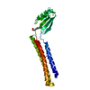
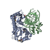
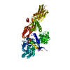
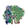
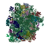
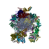
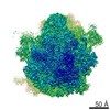
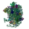
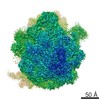
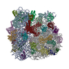
| Title | 50S subunit with EF-G(GDPNP) and RRF bound | ||||||
 Components Components |
| ||||||
 Keywords Keywords |  RIBOSOME / elongation factor G / EF-G / RRF / ribosome recycling factor / GDPNP / 50S subunit / cryo-EM / Real-space refinement / Ribonucleoprotein / Ribosomal protein / RNA-binding / rRNA-binding / Methylation / Antibiotic resistance / Repressor / Transcription / Transcription regulation / Transcription termination / Translation regulation / tRNA-binding / Phosphoprotein / Metal-binding / GTP-binding / Nucleotide-binding / Protein biosynthesis RIBOSOME / elongation factor G / EF-G / RRF / ribosome recycling factor / GDPNP / 50S subunit / cryo-EM / Real-space refinement / Ribonucleoprotein / Ribosomal protein / RNA-binding / rRNA-binding / Methylation / Antibiotic resistance / Repressor / Transcription / Transcription regulation / Transcription termination / Translation regulation / tRNA-binding / Phosphoprotein / Metal-binding / GTP-binding / Nucleotide-binding / Protein biosynthesis | ||||||
| Function / homology |  Function and homology information Function and homology informationcytoplasmic translational termination / ribosome disassembly / negative regulation of cytoplasmic translational initiation / guanosine tetraphosphate binding / translational elongation / stringent response / ribosomal large subunit binding / transcriptional attenuation / endoribonuclease inhibitor activity / RNA-binding transcription regulator activity ...cytoplasmic translational termination / ribosome disassembly / negative regulation of cytoplasmic translational initiation / guanosine tetraphosphate binding / translational elongation / stringent response / ribosomal large subunit binding / transcriptional attenuation / endoribonuclease inhibitor activity / RNA-binding transcription regulator activity / positive regulation of ribosome biogenesis / negative regulation of cytoplasmic translation / translational termination / DnaA-L2 complex / translation repressor activity / negative regulation of translational initiation / translation elongation factor activity / translational initiation / negative regulation of DNA-templated DNA replication initiation / ribosome assembly / mRNA regulatory element binding translation repressor activity / response to reactive oxygen species / assembly of large subunit precursor of preribosome / : / cytosolic ribosome assembly / regulation of cell growth / DNA-templated transcription termination / Hydrolases; Acting on acid anhydrides; Acting on GTP to facilitate cellular and subcellular movement / response to radiation / mRNA 5'-UTR binding / ribosomal large subunit assembly / large ribosomal subunit rRNA binding / ribosome binding / large ribosomal subunit / 5S rRNA binding / cytoplasmic translation / cytosolic large ribosomal subunit / transferase activity / negative regulation of translation / tRNA binding / rRNA binding / ribosome / structural constituent of ribosome / translation / response to antibiotic / mRNA binding / GTPase activity / negative regulation of DNA-templated transcription / GTP binding / DNA binding / RNA binding / zinc ion binding / cytosol / cytoplasmSimilarity search - Function | ||||||
| Biological species |  Escherichia coli (E. coli) Escherichia coli (E. coli) | ||||||
| Method | ELECTRON MICROSCOPY / single particle reconstruction / cryo EM / Resolution: 9.1 Å | ||||||
 Authors Authors | Gao, N. / Zavialov, A.V. / Ehrenberg, M. / Frank, J. | ||||||
 Citation Citation | Journal: J Mol Biol / Year: 2007 Title: Specific interaction between EF-G and RRF and its implication for GTP-dependent ribosome splitting into subunits. Authors: Ning Gao / Andrey V Zavialov / Måns Ehrenberg / Joachim Frank /  Abstract: After termination of protein synthesis, the bacterial ribosome is split into its 30S and 50S subunits by the action of ribosome recycling factor (RRF) and elongation factor G (EF-G) in a guanosine 5'- ...After termination of protein synthesis, the bacterial ribosome is split into its 30S and 50S subunits by the action of ribosome recycling factor (RRF) and elongation factor G (EF-G) in a guanosine 5'-triphosphate (GTP)-hydrolysis-dependent manner. Based on a previous cryo-electron microscopy study of ribosomal complexes, we have proposed that the binding of EF-G to an RRF-containing posttermination ribosome triggers an interdomain rotation of RRF, which destabilizes two strong intersubunit bridges (B2a and B3) and, ultimately, separates the two subunits. Here, we present a 9-A (Fourier shell correlation cutoff of 0.5) cryo-electron microscopy map of a 50S x EF-G x guanosine 5'-[(betagamma)-imido]triphosphate x RRF complex and a quasi-atomic model derived from it, showing the interaction between EF-G and RRF on the 50S subunit in the presence of the noncleavable GTP analogue guanosine 5'-[(betagamma)-imido]triphosphate. The detailed information in this model and a comparative analysis of EF-G structures in various nucleotide- and ribosome-bound states show how rotation of the RRF head domain may be triggered by various domains of EF-G. For validation of our structural model, all known mutations in EF-G and RRF that relate to ribosome recycling have been taken into account. More importantly, our results indicate a substantial conformational change in the Switch I region of EF-G, suggesting that a conformational signal transduction mechanism, similar to that employed in transfer RNA translocation on the ribosome by EF-G, translates a large-scale movement of EF-G's domain IV, induced by GTP hydrolysis, into the domain rotation of RRF that eventually splits the ribosome into subunits. #1: Journal: J.Mol.Biol. / Year: 2005Title: Structural insights into fusidic acid resistance and sensitivity in EF-G. Authors: Hansson, S. / Singh, R. / Gudkov, A.T. / Liljas, A. / Logan, D.T. #2: Journal: Science / Year: 2005Title: Structures of the bacterial ribosome at 3.5 A resolution. Authors: Schuwirth, B.S. / Borovinskaya, M.A. / Hau, C.W. / Zhang, W. / Vila-Sanjurjo, A. / Holton, J.M. / Cate, J.H. #3: Journal: Embo J. / Year: 2000Title: Crystal structure of the ribosome recycling factor from Escherichia coli. Authors: Kim, K.K. / Min, K. / Suh, S.W. #4: Journal: Nat.Struct.Mol.Biol. / Year: 2003Title: Structure of the L1 protuberance in the ribosome. Authors: Nikulin, A. / Eliseikina, I. / Tishchenko, S. / Nevskaya, N. / Davydova, N. / Platonova, O. / Piendl, W. / Selmer, M. / Liljas, A. / Drygin, D. / Zimmermann, R. / Garber, M. / Nikonov, S. #5: Journal: Mol.Cell / Year: 2005Title: Mechanism for the disassembly of the posttermination complex inferred from cryo-EM studies. Authors: Gao, N. / Zavialov, A.V. / Li, W. / Sengupta, J. / Valle, M. / Gursky, R.P. / Ehrenberg, M. / Frank, J. | ||||||
| History |
|
- Structure visualization
| Movie |
Movie viewer |
|---|---|
| Structure viewer | Molecule: MolmilJmol/JSmol |
- Downloads & links
-Download
| PDBx/mmCIF format | 2rdo.cif.gz | 256.4 KB | Display | PDBx/mmCIF format |
|---|---|---|---|---|
| PDB format | pdb2rdo.ent.gz | 144.7 KB | Display | PDB format |
| PDBx/mmJSON format | 2rdo.json.gz | Tree view | PDBx/mmJSON format | |
| Others |  Other downloads Other downloads |
-Validation report
| Arichive directory | https://data.pdbj.org/pub/pdb/validation_reports/rd/2rdoftp://data.pdbj.org/pub/pdb/validation_reports/rd/2rdo | HTTPS FTP |
|---|
-Related structure data
| Related structure data | 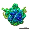 1430MC M: map data used to model this data C: citing same article ( |
|---|---|
| Similar structure data |
-Links
 PDBj
PDBj


- Assembly
| Deposited unit | 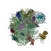
|
|---|---|
| 1 |
|
-Components
-RNA chain , 2 types, 2 molecules AB
| #1: RNA chain | / Coordinate model: P atoms only Mass: 38790.090 Da / Num. of mol.: 1 / Source method: isolated from a natural source / Source: (natural) Escherichia coli (E. coli) / References: GenBank: 33357928 |
|---|---|
| #2: RNA chain | / Coordinate model: P atoms only Mass: 941612.375 Da / Num. of mol.: 1 / Source method: isolated from a natural source / Source: (natural) Escherichia coli (E. coli) / References: GenBank: 33357927 |
+50S ribosomal protein ... , 30 types, 30 molecules VCDEFGJKLMNOPQRSTUWXYZ01234IH9
-Protein , 2 types, 2 molecules 78
| #33: Protein | EF-G / EF-G / Coordinate model: Cα atoms only Mass: 77676.227 Da / Num. of mol.: 1 / Source method: isolated from a natural source / Source: (natural) Escherichia coli (E. coli) / References: UniProt: P0A6M8 |
|---|---|
| #34: Protein | / Ribosome-releasing factor / RRF / Coordinate model: Cα atoms only Mass: 20671.621 Da / Num. of mol.: 1 / Source method: isolated from a natural source / Source: (natural) Escherichia coli (E. coli) / References: UniProt: P0A805 |
-Experimental details
-Experiment
| Experiment | Method: ELECTRON MICROSCOPY |
|---|---|
| EM experiment | Aggregation state: PARTICLE / 3D reconstruction method: single particle reconstruction |
- Sample preparation
| Component | Name: E. coli 50S complex / Type: RIBOSOME |
|---|---|
| Buffer solution | Name: PolyMix / pH: 7.5 / Details: PolyMix |
| Specimen | Conc.: 32 mg/ml / Embedding applied: NO / Shadowing applied: NO / Staining applied: NO / Vitrification applied: YES |
| Specimen support | Details: Quantifoil holey carbon film grids |
| Vitrification | Instrument: HOMEMADE PLUNGER / Cryogen name: ETHANE / Details: Rapid-freezing in liquid ethane by Vitrobot |
- Electron microscopy imaging
| Experimental equipment |  Model: Tecnai F20 / Image courtesy: FEI Company |
|---|---|
| Microscopy | Model: FEI TECNAI F20 / Date: Jan 1, 2005 |
| Electron gun | Electron source: FIELD EMISSION GUN / Accelerating voltage: 200 kV / Illumination mode: FLOOD BEAM |
| Electron lens | Mode: BRIGHT FIELDBright-field microscopy / Nominal magnification: 50000 X / Calibrated magnification: 49700 X / Nominal defocus max: 4900 nm / Nominal defocus min: 1100 nm / Cs: 2 mm |
| Specimen holder | Temperature: 93 K / Tilt angle max: 0 ° / Tilt angle min: 0 ° |
| Image recording | Electron dose: 15 e/Å2 / Film or detector model: KODAK SO-163 FILM |
| Radiation | Protocol: SINGLE WAVELENGTH / Monochromatic (M) / Laue (L): M / Scattering type: x-ray |
| Radiation wavelength | Relative weight: 1 |
- Processing
| EM software |
| |||||||||||||||||||||
|---|---|---|---|---|---|---|---|---|---|---|---|---|---|---|---|---|---|---|---|---|---|---|
| CTF correction | Details: CTF correction of 3D maps by Wiener filtration | |||||||||||||||||||||
| Symmetry | Point symmetry: C1 (asymmetric) | |||||||||||||||||||||
| 3D reconstruction | Method: 3D projection matching / Resolution: 9.1 Å / Num. of particles: 113355 / Nominal pixel size: 2.82 Å / Details: Spider Package / Symmetry type: POINT | |||||||||||||||||||||
| Atomic model building | Protocol: OTHER / Space: REAL / Details: REFINEMENT PROTOCOL--Real-space refinement | |||||||||||||||||||||
| Atomic model building |
| |||||||||||||||||||||
| Refinement step | Cycle: LAST
|

