 Movie
Movie Controller


+ Open data

- Basic information
| Entry | Database: PDB / ID: 2qx7 | ||||||
|---|---|---|---|---|---|---|---|
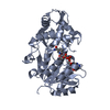
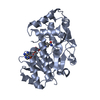
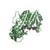
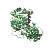
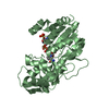
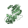
| Title | Structure of Eugenol Synthase from Ocimum basilicum | ||||||
 Components Components | Eugenol synthase 1 | ||||||
 Keywords Keywords | PLANT PROTEIN / eugenol / phenylpropene / PIP reductase / short-chain dehydrogenase/reductase | ||||||
| Function / homology |  Function and homology informationeugenol synthase / eugenol biosynthetic process / oxidoreductase activity / nucleotide binding / protein homodimerization activity Function and homology informationeugenol synthase / eugenol biosynthetic process / oxidoreductase activity / nucleotide binding / protein homodimerization activitySimilarity search - Function | ||||||
| Biological species |  Ocimum basilicum (sweet basil) Ocimum basilicum (sweet basil) | ||||||
| Method | X-RAY DIFFRACTION / SYNCHROTRON / MOLECULAR REPLACEMENT / molecular replacement / Resolution: 1.75 Å | ||||||
 Authors Authors | Louie, G.V. / Noel, J.P. / Bowman, M.E. | ||||||
 Citation Citation | Journal: Plos One / Year: 2007 Title: Structure and reaction mechanism of basil eugenol synthase Authors: Louie, G.V. / Baiga, T.J. / Bowman, M.E. / Koeduka, T. / Taylor, J.H. / Spassova, S.M. / Pichersky, E. / Noel, J.P. | ||||||
| History |
|
- Structure visualization
| Structure viewer | Molecule: MolmilJmol/JSmol |
|---|
- Downloads & links
-Download
| PDBx/mmCIF format | 2qx7.cif.gz | 147 KB | Display | PDBx/mmCIF format |
|---|---|---|---|---|
| PDB format | pdb2qx7.ent.gz | 115.2 KB | Display | PDB format |
| PDBx/mmJSON format | 2qx7.json.gz | Tree view | PDBx/mmJSON format | |
| Others |  Other downloads Other downloads |
-Validation report
| Arichive directory | https://data.pdbj.org/pub/pdb/validation_reports/qx/2qx7ftp://data.pdbj.org/pub/pdb/validation_reports/qx/2qx7 | HTTPS FTP |
|---|
-Related structure data
| Related structure data |  2qw8C  2qysC  2qzzC  2r2gC  2r6jC  1qycS C: citing same article ( S: Starting model for refinement |
|---|---|
| Similar structure data |


-Links
 PDBj
PDBj- Assembly
| Deposited unit | 
| ||||||||
|---|---|---|---|---|---|---|---|---|---|
| 1 | 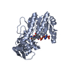
| ||||||||
| 2 | 
| ||||||||
| Unit cell |
| ||||||||
| Details | biological unit is a monomer (half of the asymmetric unit) |
-Components
| #1: Protein | Mass: 35989.465 Da / Num. of mol.: 2 Source method: isolated from a genetically manipulated source Source: (gene. exp.) Ocimum basilicum (sweet basil) / Gene: EGS1 / Plasmid: pHIS8 / Species (production host): Escherichia coli / Production host:  Escherichia coli BL21(DE3) (bacteria) / Strain (production host): BL21(DE3) / References: UniProt: Q15GI4 Escherichia coli BL21(DE3) (bacteria) / Strain (production host): BL21(DE3) / References: UniProt: Q15GI4#2: Chemical | Nicotinamide adenine dinucleotide phosphate  Mass: 743.405 Da / Num. of mol.: 2 / Source method: obtained synthetically / Formula: C21H28N7O17P3 Mass: 743.405 Da / Num. of mol.: 2 / Source method: obtained synthetically / Formula: C21H28N7O17P3#3: Water | ChemComp-HOH / | Water Mass: 18.015 Da / Num. of mol.: 547 / Source method: isolated from a natural source / Formula: H2O Mass: 18.015 Da / Num. of mol.: 547 / Source method: isolated from a natural source / Formula: H2O |
|---|
-Experimental details
-Experiment
| Experiment | Method: X-RAY DIFFRACTION / Number of used crystals: 1 |
|---|
- Sample preparation
| Crystal | Density Matthews: 2.37 Å3/Da / Density % sol: 48.11 % |
|---|---|
| Crystal grow | Temperature: 277 K / Method: vapor diffusion, hanging drop / pH: 5.5 Details: 0.1 M sodium succinate, 21% PEG 3350, 0.3 M KCl, 2 mM dithiothreitol, 5 mM NADP+, pH 5.5, vapor diffusion, hanging drop, temperature 277K |
-Data collection
| Diffraction | Mean temperature: 100 K |
|---|---|
| Diffraction source | Source: SYNCHROTRON / Site: ALS  / Beamline: 8.2.2 / Wavelength: 1 Å / Beamline: 8.2.2 / Wavelength: 1 Å |
| Detector | Type: ADSC QUANTUM 315 / Detector: CCD / Date: Dec 1, 2005 |
| Radiation | Monochromator: double crystal Si(111) / Protocol: SINGLE WAVELENGTH / Scattering type: x-ray |
| Radiation wavelength | Wavelength: 1 Å / Relative weight: 1 |
| Reflection | Highest resolution: 1.75 Å / Num. obs: 68896 / % possible obs: 99.9 % / Observed criterion σ(I): -3 / Redundancy: 6.99 % / Biso Wilson estimate: 26.18 Å2 / Rmerge(I) obs: 0.105 / Net I/σ(I): 10.54 |
| Reflection shell | Resolution: 1.75→1.84 Å / Redundancy: 5.83 % / Rmerge(I) obs: 0.662 / Mean I/σ(I) obs: 3 / Num. measured obs: 55088 / Num. unique obs: 9455 / % possible all: 99.3 |
-Phasing
| Phasing | Method: molecular replacement | |||||||||
|---|---|---|---|---|---|---|---|---|---|---|
| Phasing MR | Rfactor: 0.568 / Cor.coef. Fo:Fc: 0.495
|
- Processing
| Software |
| ||||||||||||||||||||||||||||
|---|---|---|---|---|---|---|---|---|---|---|---|---|---|---|---|---|---|---|---|---|---|---|---|---|---|---|---|---|---|
| Refinement | Method to determine structure: MOLECULAR REPLACEMENT Starting model: PDB entry 1QYC Resolution: 1.75→42.05 Å / FOM work R set: 0.744 / Isotropic thermal model: Isotropic / Cross valid method: THROUGHOUT / σ(F): 0 / Stereochemistry target values: Engh & Huber
| ||||||||||||||||||||||||||||
| Solvent computation | Bsol: 50.005 Å2 | ||||||||||||||||||||||||||||
| Displacement parameters | Biso mean: 24.074 Å2
| ||||||||||||||||||||||||||||
| Refinement step | Cycle: LAST / Resolution: 1.75→42.05 Å
| ||||||||||||||||||||||||||||
| Refine LS restraints |
| ||||||||||||||||||||||||||||
| Xplor file |
|
