 Movie
Movie Controller


+ Open data

- Basic information
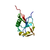
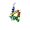
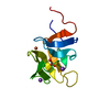
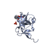
| Entry | Database: PDB / ID: 2qvo | ||||||
|---|---|---|---|---|---|---|---|
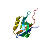
| Title | Crystal structure of AF1382 from Archaeoglobus fulgidus | ||||||
 Components Components | Uncharacterized protein AF_1382 | ||||||
 Keywords Keywords |  STRUCTURAL GENOMICS / UNKNOWN FUNCTION / AF1382 / Archaeoglobus fulgidus / PSI / SOUTHEAST COLLABORATORY FOR STRUCTURAL GENOMICS / Protein Structure Initiative / SECSG STRUCTURAL GENOMICS / UNKNOWN FUNCTION / AF1382 / Archaeoglobus fulgidus / PSI / SOUTHEAST COLLABORATORY FOR STRUCTURAL GENOMICS / Protein Structure Initiative / SECSG | ||||||
| Function / homology | Winged helix-like DNA-binding domain superfamily/Winged helix DNA-binding domain / Arc Repressor Mutant, subunit A / Winged helix DNA-binding domain superfamily / Winged helix-like DNA-binding domain superfamily / Orthogonal Bundle / Mainly Alpha / Uncharacterized protein AF_1382 Function and homology information Function and homology information | ||||||
| Biological species |   Archaeoglobus fulgidus DSM 4304 (archaea) Archaeoglobus fulgidus DSM 4304 (archaea) | ||||||
| Method | X-RAY DIFFRACTION / SYNCHROTRON / Sulfur SAD / Resolution: 1.85 Å | ||||||
 Authors Authors | Zhu, J. / Zhao, M. / Fu, Z.-Q. / Yang, H. / Chang, J. / Hao, X. / Chen, L. / Liu, Z.J. / Rose, J.P. / Wang, B.C. / Southeast Collaboratory for Structural Genomics (SECSG) | ||||||
 Citation Citation | Journal: To be Published Title: Crystal structure of AF1382 from Archaeoglobus fulgidus. Authors: Zhu, J. / Zhao, M. / Fu, Z.-Q. / Yang, H. / Chang, J. / Hao, X. / Chen, L. / Liu, Z.J. / Rose, J.P. / Wang, B.C. | ||||||
| History |
|
- Structure visualization
| Structure viewer | Molecule: MolmilJmol/JSmol |
|---|
- Downloads & links
-Download
| PDBx/mmCIF format | 2qvo.cif.gz | 31.7 KB | Display | PDBx/mmCIF format |
|---|---|---|---|---|
| PDB format | pdb2qvo.ent.gz | 20.8 KB | Display | PDB format |
| PDBx/mmJSON format | 2qvo.json.gz | Tree view | PDBx/mmJSON format | |
| Others |  Other downloads Other downloads |
-Validation report
| Arichive directory | https://data.pdbj.org/pub/pdb/validation_reports/qv/2qvoftp://data.pdbj.org/pub/pdb/validation_reports/qv/2qvo | HTTPS FTP |
|---|
-Related structure data
| Similar structure data |
|---|


-Links
 PDBj
PDBj- Assembly
| Deposited unit | 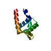
| ||||||||
|---|---|---|---|---|---|---|---|---|---|
| 1 |
| ||||||||
| 2 | 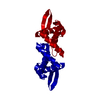
| ||||||||
| Unit cell |
| ||||||||
| Components on special symmetry positions |
|
-Components
| #1: Protein | Mass: 11159.016 Da / Num. of mol.: 1 Source method: isolated from a genetically manipulated source Source: (gene. exp.) Archaeoglobus fulgidus DSM 4304 (archaea)Species: Archaeoglobus fulgidus / Strain: VC-16, JCM 9628, NBRC 100126 / Gene: AF_1382 / Plasmid: pDEST-527 / Production host:  Escherichia coli (E. coli) / Strain (production host): BL21(DE3)-RPX / References: UniProt: O28889 Escherichia coli (E. coli) / Strain (production host): BL21(DE3)-RPX / References: UniProt: O28889 |
|---|---|
| #2: Water | ChemComp-HOH / Water Mass: 18.015 Da / Num. of mol.: 38 / Source method: isolated from a natural source / Formula: H2O Mass: 18.015 Da / Num. of mol.: 38 / Source method: isolated from a natural source / Formula: H2O |
-Experimental details
-Experiment
| Experiment | Method: X-RAY DIFFRACTION / Number of used crystals: 1 |
|---|
- Sample preparation
| Crystal | Density Matthews: 2.7 Å3/Da / Density % sol: 54.4 % |
|---|---|
| Crystal grow | Temperature: 291 K / Method: microbatch / pH: 4.6 Details: USING 1.0 MICROLITER DROPS CONTAINING EQUAL VOLUMES OF PROTEIN CONCENTRATE (2.8 mg/ml) AND SOLUTION CONTAINING 0.2 M AMMONIUM SULFATE, 0.1 M SODIUM ACETATE, 30% w/v POLYETHYLENE GLYCOL ...Details: USING 1.0 MICROLITER DROPS CONTAINING EQUAL VOLUMES OF PROTEIN CONCENTRATE (2.8 mg/ml) AND SOLUTION CONTAINING 0.2 M AMMONIUM SULFATE, 0.1 M SODIUM ACETATE, 30% w/v POLYETHYLENE GLYCOL MONOMETHYL ETHER, pH 4.6, MICROBATCH, temperature 291K |
-Data collection
| Diffraction | Mean temperature: 100 K | |||||||||
|---|---|---|---|---|---|---|---|---|---|---|
| Diffraction source | Source: SYNCHROTRON / Site: APS  / Beamline: 22-ID / Wavelength: 0.97240, 1.90000 / Beamline: 22-ID / Wavelength: 0.97240, 1.90000 | |||||||||
| Detector | Type: MARMOSAIC 300 mm CCD / Detector: CCD / Date: Aug 1, 2007 / Details: ROSENBAUM | |||||||||
| Radiation | Monochromator: SI CHANNEL 220 / Protocol: SINGLE WAVELENGTH / Monochromatic (M) / Laue (L): M / Scattering type: x-ray | |||||||||
| Radiation wavelength |
| |||||||||
| Reflection | Resolution: 1.85→50 Å / Num. obs: 19060 / % possible obs: 99.9 % / Redundancy: 13.3 % / Rsym value: 0.051 / Net I/σ(I): 12.96 | |||||||||
| Reflection shell | Resolution: 1.85→1.92 Å / Redundancy: 12.5 % / Rsym value: 0.233 / % possible all: 99.8 |
- Processing
| Software |
| ||||||||||||||||||||||||||||||||||||||||||||||||||||||||||||||||||||||||||||||||||||||||||||||||||||||||||||||||||||||||||||||||||||||||||||||||||||||||||||||||||||||||||
|---|---|---|---|---|---|---|---|---|---|---|---|---|---|---|---|---|---|---|---|---|---|---|---|---|---|---|---|---|---|---|---|---|---|---|---|---|---|---|---|---|---|---|---|---|---|---|---|---|---|---|---|---|---|---|---|---|---|---|---|---|---|---|---|---|---|---|---|---|---|---|---|---|---|---|---|---|---|---|---|---|---|---|---|---|---|---|---|---|---|---|---|---|---|---|---|---|---|---|---|---|---|---|---|---|---|---|---|---|---|---|---|---|---|---|---|---|---|---|---|---|---|---|---|---|---|---|---|---|---|---|---|---|---|---|---|---|---|---|---|---|---|---|---|---|---|---|---|---|---|---|---|---|---|---|---|---|---|---|---|---|---|---|---|---|---|---|---|---|---|---|---|
| Refinement | Method to determine structure: Sulfur SAD / Resolution: 1.85→37.5 Å / Cor.coef. Fo:Fc: 0.93 / Cor.coef. Fo:Fc free: 0.897 / Cross valid method: THROUGHOUT / ESU R: 0.15 / ESU R Free: 0.147 / Stereochemistry target values: MAXIMUM LIKELIHOOD Details: 1. Initial phases were obtained from sulfur phasing using data collected at wavelength 1.90000 A. Then the structure was refined using 0.9724 A - wavelength data and deposited in PDB. 2. The ...Details: 1. Initial phases were obtained from sulfur phasing using data collected at wavelength 1.90000 A. Then the structure was refined using 0.9724 A - wavelength data and deposited in PDB. 2. The Bijvoet differences were used for phasing. 3. HYDROGENS HAVE BEEN ADDED IN THE RIDING POSITIONS.
| ||||||||||||||||||||||||||||||||||||||||||||||||||||||||||||||||||||||||||||||||||||||||||||||||||||||||||||||||||||||||||||||||||||||||||||||||||||||||||||||||||||||||||
| Solvent computation | Ion probe radii: 0.8 Å / Shrinkage radii: 0.8 Å / VDW probe radii: 1.2 Å / Solvent model: MASK | ||||||||||||||||||||||||||||||||||||||||||||||||||||||||||||||||||||||||||||||||||||||||||||||||||||||||||||||||||||||||||||||||||||||||||||||||||||||||||||||||||||||||||
| Displacement parameters | Biso mean: 23.008 Å2
| ||||||||||||||||||||||||||||||||||||||||||||||||||||||||||||||||||||||||||||||||||||||||||||||||||||||||||||||||||||||||||||||||||||||||||||||||||||||||||||||||||||||||||
| Refinement step | Cycle: LAST / Resolution: 1.85→37.5 Å
| ||||||||||||||||||||||||||||||||||||||||||||||||||||||||||||||||||||||||||||||||||||||||||||||||||||||||||||||||||||||||||||||||||||||||||||||||||||||||||||||||||||||||||
| Refine LS restraints |
| ||||||||||||||||||||||||||||||||||||||||||||||||||||||||||||||||||||||||||||||||||||||||||||||||||||||||||||||||||||||||||||||||||||||||||||||||||||||||||||||||||||||||||
| LS refinement shell | Resolution: 1.85→1.898 Å / Total num. of bins used: 20
|
