 Movie
Movie Controller

[English] 日本語
 Yorodumi
- PDB-2qlw: Crystal structure of rhamnose mutarotase RhaU of Rhizobium legumi... -
+ Open data

- Basic information
| Entry | Database: PDB / ID: 2qlw | ||||||
|---|---|---|---|---|---|---|---|
| Title | Crystal structure of rhamnose mutarotase RhaU of Rhizobium leguminosarum | ||||||
 Components Components | RhaU | ||||||
 Keywords Keywords | ISOMERASE / RhaU / mutarotase / Rhizobium leguminosarum | ||||||
| Function / homology |  Function and homology information Function and homology informationL-rhamnose mutarotase / L-rhamnose mutarotase activity / rhamnose catabolic process / cytoplasmSimilarity search - Function | ||||||
| Biological species |  Rhizobium leguminosarum bv. trifolii (bacteria) Rhizobium leguminosarum bv. trifolii (bacteria) | ||||||
| Method | X-RAY DIFFRACTION / SYNCHROTRON / SAD / Resolution: 1.6 Å | ||||||
 Authors Authors | Carpena, X. / Loewen, P.C. | ||||||
 Citation Citation | Journal: J.Bacteriol. / Year: 2008 Title: RhaU of Rhizobium leguminosarum is a rhamnose mutarotase. Authors: Richardson, J.S. / Carpena, X. / Switala, J. / Perez-Luque, R. / Donald, L.J. / Loewen, P.C. / Oresnik, I.J. | ||||||
| History |
|
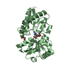
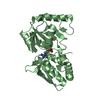
- Structure visualization
| Structure viewer | Molecule: MolmilJmol/JSmol |
|---|
- Downloads & links
-Download
| PDBx/mmCIF format | 2qlw.cif.gz | 65.8 KB | Display | PDBx/mmCIF format |
|---|---|---|---|---|
| PDB format | pdb2qlw.ent.gz | 52.5 KB | Display | PDB format |
| PDBx/mmJSON format | 2qlw.json.gz | Tree view | PDBx/mmJSON format | |
| Others |  Other downloads Other downloads |
-Validation report
| Arichive directory | https://data.pdbj.org/pub/pdb/validation_reports/ql/2qlwftp://data.pdbj.org/pub/pdb/validation_reports/ql/2qlw | HTTPS FTP |
|---|
-Related structure data


-Links
 PDBj
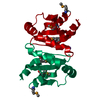
PDBj- Assembly
| Deposited unit | 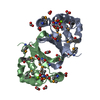
| ||||||||||||||||||
|---|---|---|---|---|---|---|---|---|---|---|---|---|---|---|---|---|---|---|---|
| 1 |
| ||||||||||||||||||
| 2 | 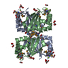
| ||||||||||||||||||
| Unit cell |
| ||||||||||||||||||
| Noncrystallographic symmetry (NCS) | NCS domain:
NCS domain segments: Component-ID: 1 / Ens-ID: 1 / Beg label comp-ID: MSE / End label comp-ID: PHE / Refine code: 6 / Auth seq-ID: 1 - 103 / Label seq-ID: 3 - 105
|
-Components
| #1: Protein | Mass: 16996.836 Da / Num. of mol.: 2 Source method: isolated from a genetically manipulated source Source: (gene. exp.) Rhizobium leguminosarum bv. trifolii (bacteria)Gene: rhaU / Production host: Escherichia coli (E. coli) / References: UniProt: Q7BSH1#2: Chemical |   Mass: 24.305 Da / Num. of mol.: 2 / Source method: obtained synthetically / Formula: Mg Mass: 24.305 Da / Num. of mol.: 2 / Source method: obtained synthetically / Formula: Mg#3: Chemical | ChemComp-FMT / Formic acid  Mass: 46.025 Da / Num. of mol.: 16 / Source method: obtained synthetically / Formula: CH2O2 Mass: 46.025 Da / Num. of mol.: 16 / Source method: obtained synthetically / Formula: CH2O2#4: Water | ChemComp-HOH / | Water Mass: 18.015 Da / Num. of mol.: 220 / Source method: isolated from a natural source / Formula: H2O Mass: 18.015 Da / Num. of mol.: 220 / Source method: isolated from a natural source / Formula: H2O |
|---|
-Experimental details
-Experiment
| Experiment | Method: X-RAY DIFFRACTION / Number of used crystals: 1 |
|---|
- Sample preparation
| Crystal | Density Matthews: 2.72 Å3/Da / Density % sol: 54.76 % |
|---|---|
| Crystal grow | Temperature: 298 K / Method: vapor diffusion, hanging drop / pH: 4.6 Details: 2M Sodium Formate, 0.1M Sodium Acetate pH 4.6, 20% Glycerol , VAPOR DIFFUSION, HANGING DROP, temperature 298K |
-Data collection
| Diffraction | Mean temperature: 100 K | |||||||||||||||||||||||||||||||||||||||||||||||||||||||||||||||||||||||||||||
|---|---|---|---|---|---|---|---|---|---|---|---|---|---|---|---|---|---|---|---|---|---|---|---|---|---|---|---|---|---|---|---|---|---|---|---|---|---|---|---|---|---|---|---|---|---|---|---|---|---|---|---|---|---|---|---|---|---|---|---|---|---|---|---|---|---|---|---|---|---|---|---|---|---|---|---|---|---|---|
| Diffraction source | Source: SYNCHROTRON / Site: ESRF  / Beamline: BM16 / Wavelength: 0.9079 Å / Beamline: BM16 / Wavelength: 0.9079 Å | |||||||||||||||||||||||||||||||||||||||||||||||||||||||||||||||||||||||||||||
| Detector | Type: MAR CCD 165 mm / Detector: CCD / Date: Feb 23, 2006 / Details: Mirrors | |||||||||||||||||||||||||||||||||||||||||||||||||||||||||||||||||||||||||||||
| Radiation | Monochromator: SI 111 / Protocol: MAD / Monochromatic (M) / Laue (L): M / Scattering type: x-ray | |||||||||||||||||||||||||||||||||||||||||||||||||||||||||||||||||||||||||||||
| Radiation wavelength | Wavelength: 0.9079 Å / Relative weight: 1 | |||||||||||||||||||||||||||||||||||||||||||||||||||||||||||||||||||||||||||||
| Reflection | Redundancy: 5.7 % / Av σ(I) over netI: 11.2 / Number: 203502 / Rmerge(I) obs: 0.097 / Χ2: 0.95 / D res high: 1.6 Å / D res low: 30 Å / Num. obs: 35978 / % possible obs: 97.1 | |||||||||||||||||||||||||||||||||||||||||||||||||||||||||||||||||||||||||||||
| Diffraction reflection shell |
| |||||||||||||||||||||||||||||||||||||||||||||||||||||||||||||||||||||||||||||
| Reflection | Resolution: 1.6→30 Å / Num. all: 35978 / Num. obs: 35978 / % possible obs: 97.1 % / Observed criterion σ(F): 0 / Observed criterion σ(I): 0 / Redundancy: 5.7 % / Rmerge(I) obs: 0.097 / Χ2: 0.954 / Net I/σ(I): 11.2 | |||||||||||||||||||||||||||||||||||||||||||||||||||||||||||||||||||||||||||||
| Reflection shell | Resolution: 1.6→1.66 Å / Redundancy: 5.6 % / Rmerge(I) obs: 0.589 / Mean I/σ(I) obs: 3.2 / Num. unique all: 3574 / Χ2: 0.98 / % possible all: 97 |
-Phasing
| Phasing | Method: SAD |
|---|
- Processing
| Software |
| |||||||||||||||||||||||||||||||||||||||||||||||||||||||||||||||||||||||||||||||||||||||||||||||||||||||||||||||||||||||||||||
|---|---|---|---|---|---|---|---|---|---|---|---|---|---|---|---|---|---|---|---|---|---|---|---|---|---|---|---|---|---|---|---|---|---|---|---|---|---|---|---|---|---|---|---|---|---|---|---|---|---|---|---|---|---|---|---|---|---|---|---|---|---|---|---|---|---|---|---|---|---|---|---|---|---|---|---|---|---|---|---|---|---|---|---|---|---|---|---|---|---|---|---|---|---|---|---|---|---|---|---|---|---|---|---|---|---|---|---|---|---|---|---|---|---|---|---|---|---|---|---|---|---|---|---|---|---|---|
| Refinement | Method to determine structure: SAD / Resolution: 1.6→19.99 Å / Cor.coef. Fo:Fc: 0.966 / Cor.coef. Fo:Fc free: 0.954 / SU B: 2.496 / SU ML: 0.044 / TLS residual ADP flag: LIKELY RESIDUAL / Cross valid method: THROUGHOUT / σ(F): 0 / σ(I): 0 / ESU R: 0.072 / ESU R Free: 0.074 / Stereochemistry target values: MAXIMUM LIKELIHOOD / Details: HYDROGENS HAVE BEEN ADDED IN THE RIDING POSITIONS
| |||||||||||||||||||||||||||||||||||||||||||||||||||||||||||||||||||||||||||||||||||||||||||||||||||||||||||||||||||||||||||||
| Solvent computation | Ion probe radii: 0.8 Å / Shrinkage radii: 0.8 Å / VDW probe radii: 1.2 Å / Solvent model: MASK | |||||||||||||||||||||||||||||||||||||||||||||||||||||||||||||||||||||||||||||||||||||||||||||||||||||||||||||||||||||||||||||
| Displacement parameters | Biso mean: 10.823 Å2
| |||||||||||||||||||||||||||||||||||||||||||||||||||||||||||||||||||||||||||||||||||||||||||||||||||||||||||||||||||||||||||||
| Refinement step | Cycle: LAST / Resolution: 1.6→19.99 Å
| |||||||||||||||||||||||||||||||||||||||||||||||||||||||||||||||||||||||||||||||||||||||||||||||||||||||||||||||||||||||||||||
| Refine LS restraints |
| |||||||||||||||||||||||||||||||||||||||||||||||||||||||||||||||||||||||||||||||||||||||||||||||||||||||||||||||||||||||||||||
| Refine LS restraints NCS | Dom-ID: 1 / Auth asym-ID: A / Ens-ID: 1 / Number: 1381 / Refine-ID: X-RAY DIFFRACTION
| |||||||||||||||||||||||||||||||||||||||||||||||||||||||||||||||||||||||||||||||||||||||||||||||||||||||||||||||||||||||||||||
| LS refinement shell | Resolution: 1.6→1.641 Å / Total num. of bins used: 20
| |||||||||||||||||||||||||||||||||||||||||||||||||||||||||||||||||||||||||||||||||||||||||||||||||||||||||||||||||||||||||||||
| Refinement TLS params. | Method: refined / Refine-ID: X-RAY DIFFRACTION
| |||||||||||||||||||||||||||||||||||||||||||||||||||||||||||||||||||||||||||||||||||||||||||||||||||||||||||||||||||||||||||||
| Refinement TLS group |
|
