 Movie
Movie Controller

[English] 日本語
 Yorodumi
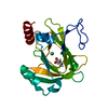
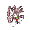
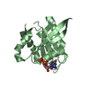
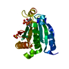
- PDB-2q3f: X-ray crystal structure of putative human Ras-related GTP binding... -
+ Open data

- Basic information
| Entry | Database: PDB / ID: 2q3f | ||||||
|---|---|---|---|---|---|---|---|
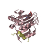
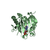
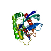
| Title | X-ray crystal structure of putative human Ras-related GTP binding D in complex with GMPPNP | ||||||
 Components Components | Ras-related GTP-binding protein D | ||||||
 Keywords Keywords |  PROTEIN BINDING / Structural Genomics / GTP-binding / RRAGD / Structural Genomics Consortium / SGC PROTEIN BINDING / Structural Genomics / GTP-binding / RRAGD / Structural Genomics Consortium / SGC | ||||||
| Function / homology |  Function and homology information Function and homology informationRagulator complex => GO:0071986 / regulation of cell cycle => GO:0051726 / protein localization => GO:0008104 / Gtr1-Gtr2 GTPase complex / cellular response to leucine starvation / cellular response to L-leucine / MTOR signalling / Amino acids regulate mTORC1 / Energy dependent regulation of mTOR by LKB1-AMPK / Macroautophagy ...Ragulator complex => GO:0071986 / regulation of cell cycle => GO:0051726 / protein localization => GO:0008104 / Gtr1-Gtr2 GTPase complex / cellular response to leucine starvation / cellular response to L-leucine / MTOR signalling / Amino acids regulate mTORC1 / Energy dependent regulation of mTOR by LKB1-AMPK / Macroautophagy / mTORC1-mediated signalling / positive regulation of TOR signaling / regulation of macroautophagy / positive regulation of TORC1 signaling / cellular response to starvation / Regulation of PTEN gene transcription / regulation of autophagy / cellular response to amino acid stimulus / TP53 Regulates Metabolic Genes / Hydrolases; Acting on acid anhydrides; Acting on GTP to facilitate cellular and subcellular movement / GDP binding / GTPase binding / lysosome / protein heterodimerization activity / GTPase activity / centrosome / GTP binding / nucleoplasm / nucleus / cytosol / cytoplasmSimilarity search - Function | ||||||
| Biological species |  Homo sapiens (human) Homo sapiens (human) | ||||||
| Method | X-RAY DIFFRACTION / SYNCHROTRON / SAD / Resolution: 2.1 Å | ||||||
 Authors Authors | Mulichak, A.M. / Rabeh, W.M. / Tempel, W. / Nedyalkova, L. / Landry, R. / Arrowsmith, C.H. / Edwards, A.M. / Sundstrom, M. / Weigelt, J. / Keefe, L.J. ...Mulichak, A.M. / Rabeh, W.M. / Tempel, W. / Nedyalkova, L. / Landry, R. / Arrowsmith, C.H. / Edwards, A.M. / Sundstrom, M. / Weigelt, J. / Keefe, L.J. / Bochkarev, A. / Park, H. / Structural Genomics Consortium (SGC) | ||||||
 Citation Citation | Journal: To be published Title: Crystal structure of human Ras-related GTP-binding D. Authors: Mulichak, A.M. / Rabeh, W.M. / Tempel, W. / Nedyalkova, L. / Landry, R. / Arrowsmith, C.H. / Edwards, A.M. / Sundstrom, M. / Weigelt, J. / Keefe, L.J. / Bochkarev, A. / Park, H. | ||||||
| History |
|
- Structure visualization
| Structure viewer | Molecule: MolmilJmol/JSmol |
|---|
- Downloads & links
-Download
| PDBx/mmCIF format | 2q3f.cif.gz | 83 KB | Display | PDBx/mmCIF format |
|---|---|---|---|---|
| PDB format | pdb2q3f.ent.gz | 67.5 KB | Display | PDB format |
| PDBx/mmJSON format | 2q3f.json.gz | Tree view | PDBx/mmJSON format | |
| Others |  Other downloads Other downloads |
-Validation report
| Arichive directory | https://data.pdbj.org/pub/pdb/validation_reports/q3/2q3fftp://data.pdbj.org/pub/pdb/validation_reports/q3/2q3f | HTTPS FTP |
|---|
-Related structure data
| Similar structure data |
|---|


-Links
 PDBj
PDBj


- Assembly
| Deposited unit | 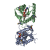
| ||||||||
|---|---|---|---|---|---|---|---|---|---|
| 1 | 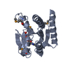
| ||||||||
| 2 | 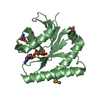
| ||||||||
| Unit cell |
| ||||||||
| Details | The biological assembly is a monomer. |
-Components
| #1: Protein | Mass: 21126.332 Da / Num. of mol.: 2 Source method: isolated from a genetically manipulated source Source: (gene. exp.) Homo sapiens (human) / Gene: RRAGD / Plasmid: p28a-LIC / Species (production host): Escherichia coli / Production host:  Escherichia coli BL21(DE3) (bacteria) / Strain (production host): BL21 (DE3) / References: UniProt: Q9NQL2 Escherichia coli BL21(DE3) (bacteria) / Strain (production host): BL21 (DE3) / References: UniProt: Q9NQL2#2: Chemical |   Mass: 24.305 Da / Num. of mol.: 2 / Source method: obtained synthetically / Formula: Mg Mass: 24.305 Da / Num. of mol.: 2 / Source method: obtained synthetically / Formula: Mg#3: Chemical | 5'-Guanylyl imidodiphosphate  Mass: 522.196 Da / Num. of mol.: 2 / Source method: obtained synthetically / Formula: C10H17N6O13P3 Mass: 522.196 Da / Num. of mol.: 2 / Source method: obtained synthetically / Formula: C10H17N6O13P3Comment: GppNHp, GMPPNP, energy-carrying molecule analogue*YM #4: Water | ChemComp-HOH / | Water Mass: 18.015 Da / Num. of mol.: 90 / Source method: isolated from a natural source / Formula: H2O Mass: 18.015 Da / Num. of mol.: 90 / Source method: isolated from a natural source / Formula: H2O |
|---|
-Experimental details
-Experiment
| Experiment | Method: X-RAY DIFFRACTION / Number of used crystals: 1 |
|---|
- Sample preparation
| Crystal | Density Matthews: 2.35 Å3/Da / Density % sol: 47.65 % |
|---|---|
| Crystal grow | Temperature: 298 K / Method: vapor diffusion / pH: 8.4 Details: 31% PEG4000, 0.2M sodium acetate and 0.1M Tris HCl, pH 8.4, VAPOR DIFFUSION, temperature 298K |
-Data collection
| Diffraction | Mean temperature: 100 K | |||||||||
|---|---|---|---|---|---|---|---|---|---|---|
| Diffraction source | Source: SYNCHROTRON / Site: APS  / Beamline: 17-ID / Wavelength: 0.97942, 1.0 / Beamline: 17-ID / Wavelength: 0.97942, 1.0 | |||||||||
| Detector | Type: ADSC QUANTUM 210 / Detector: CCD / Date: Apr 14, 2007 | |||||||||
| Radiation | Monochromator: Si 111 / Protocol: SINGLE WAVELENGTH / Monochromatic (M) / Laue (L): M / Scattering type: x-ray | |||||||||
| Radiation wavelength |
| |||||||||
| Reflection | Resolution: 2.05→50 Å / Num. all: 24803 / Num. obs: 24803 / % possible obs: 99.8 % / Observed criterion σ(F): 0 / Redundancy: 4.1 % / Rsym value: 0.054 | |||||||||
| Reflection shell | Resolution: 2.05→2.12 Å / Redundancy: 3.8 % / Rmerge(I) obs: 0.542 / Mean I/σ(I) obs: 2.4 / Num. unique all: 2438 / % possible all: 99.1 |
- Processing
| Software |
| |||||||||||||||||||||||||
|---|---|---|---|---|---|---|---|---|---|---|---|---|---|---|---|---|---|---|---|---|---|---|---|---|---|---|
| Refinement | Method to determine structure: SAD / Resolution: 2.1→50 Å / Isotropic thermal model: Isotropic / Cross valid method: THROUGHOUT / σ(F): 0 / Stereochemistry target values: Engh & Huber
| |||||||||||||||||||||||||
| Refinement step | Cycle: LAST / Resolution: 2.1→50 Å
| |||||||||||||||||||||||||
| Refine LS restraints |
|
