 Movie
Movie Controller

[English] 日本語
 Yorodumi
- PDB-2py3: Crystal Structure of FGF Receptor 2 (FGFR2) Kinase Domain Harbori... -
+ Open data

- Basic information
| Entry | Database: PDB / ID: 2py3 | ||||||
|---|---|---|---|---|---|---|---|
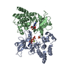
| Title | Crystal Structure of FGF Receptor 2 (FGFR2) Kinase Domain Harboring the Pathogenic E565G Mutation Responsible for Pfeiffer Syndrome | ||||||
 Components Components | Fibroblast growth factor receptor 2 | ||||||
 Keywords Keywords | TRANSFERASE / Kinase Domain Fold Consisting of N- and C-lobes | ||||||
| Function / homology |  Function and homology information Function and homology informationSignaling by FGFR2 amplification mutants / Signaling by FGFR2 fusions / fibroblast growth factor receptor signaling pathway involved in negative regulation of apoptotic process in bone marrow cell / fibroblast growth factor receptor signaling pathway involved in hemopoiesis / fibroblast growth factor receptor signaling pathway involved in positive regulation of cell proliferation in bone marrow / fibroblast growth factor receptor signaling pathway involved in mammary gland specification / lateral sprouting from an epithelium / mammary gland bud formation / branch elongation involved in salivary gland morphogenesis / mesenchymal cell differentiation involved in lung development ...Signaling by FGFR2 amplification mutants / Signaling by FGFR2 fusions / fibroblast growth factor receptor signaling pathway involved in negative regulation of apoptotic process in bone marrow cell / fibroblast growth factor receptor signaling pathway involved in hemopoiesis / fibroblast growth factor receptor signaling pathway involved in positive regulation of cell proliferation in bone marrow / fibroblast growth factor receptor signaling pathway involved in mammary gland specification / lateral sprouting from an epithelium / mammary gland bud formation / branch elongation involved in salivary gland morphogenesis / mesenchymal cell differentiation involved in lung development / lacrimal gland development / otic vesicle formation / prostate gland morphogenesis / regulation of smooth muscle cell differentiation / orbitofrontal cortex development / regulation of morphogenesis of a branching structure / squamous basal epithelial stem cell differentiation involved in prostate gland acinus development / branching morphogenesis of a nerve / embryonic organ morphogenesis / endochondral bone growth / morphogenesis of embryonic epithelium / epidermis morphogenesis / pyramidal neuron development / bud elongation involved in lung branching / positive regulation of epithelial cell proliferation involved in lung morphogenesis / membranous septum morphogenesis / reproductive structure development / limb bud formation / lung lobe morphogenesis / gland morphogenesis / fibroblast growth factor receptor signaling pathway involved in orbitofrontal cortex development / ventricular zone neuroblast division / embryonic digestive tract morphogenesis / epithelial cell proliferation involved in salivary gland morphogenesis / mesenchymal cell differentiation / mesenchymal cell proliferation involved in lung development / branching involved in prostate gland morphogenesis / branching involved in labyrinthine layer morphogenesis / FGFR2b ligand binding and activation / FGFR2c ligand binding and activation / Activated point mutants of FGFR2 / regulation of osteoblast proliferation / Phospholipase C-mediated cascade; FGFR2 / fibroblast growth factor receptor activity / branching involved in salivary gland morphogenesis / embryonic pattern specification / outflow tract septum morphogenesis / positive regulation of phospholipase activity / lung-associated mesenchyme development / mesodermal cell differentiation / digestive tract development / embryonic cranial skeleton morphogenesis / regulation of smoothened signaling pathway / bone morphogenesis / skeletal system morphogenesis / odontogenesis / ureteric bud development / positive regulation of mesenchymal cell proliferation / regulation of osteoblast differentiation / ventricular cardiac muscle tissue morphogenesis / Signaling by FGFR2 IIIa TM / inner ear morphogenesis / organ growth / midbrain development / hair follicle morphogenesis / lung alveolus development / fibroblast growth factor binding / prostate epithelial cord arborization involved in prostate glandular acinus morphogenesis / PI-3K cascade:FGFR2 / bone mineralization / prostate epithelial cord elongation / excitatory synapse / positive regulation of cell division / positive regulation of Wnt signaling pathway / PI3K Cascade / epithelial to mesenchymal transition / negative regulation of keratinocyte proliferation / fibroblast growth factor receptor signaling pathway / cell fate commitment / embryonic organ development / positive regulation of cell cycle / SHC-mediated cascade:FGFR2 / cellular response to retinoic acid / FRS-mediated FGFR2 signaling / positive regulation of cardiac muscle cell proliferation / positive regulation of vascular associated smooth muscle cell proliferation / cellular response to transforming growth factor beta stimulus / Signaling by FGFR2 in disease / epithelial cell differentiation / axonogenesis / post-embryonic development / regulation of ERK1 and ERK2 cascade / positive regulation of epithelial cell proliferation / Negative regulation of FGFR2 signaling / lung development / animal organ morphogenesis / bone development / receptor protein-tyrosine kinase / peptidyl-tyrosine phosphorylation / positive regulation of canonical Wnt signaling pathwaySimilarity search - Function | ||||||
| Biological species |  Homo sapiens (human) Homo sapiens (human) | ||||||
| Method | X-RAY DIFFRACTION / SYNCHROTRON / MOLECULAR REPLACEMENT / Resolution: 2.3 Å | ||||||
 Authors Authors | Chen, H. / Mohammadi, M. | ||||||
 Citation Citation | Journal: Mol.Cell / Year: 2007 Title: A molecular brake in the kinase hinge region regulates the activity of receptor tyrosine kinases. Authors: Chen, H. / Ma, J. / Li, W. / Eliseenkova, A.V. / Xu, C. / Neubert, T.A. / Miller, W.T. / Mohammadi, M. | ||||||
| History |
|
- Structure visualization
| Structure viewer | Molecule: MolmilJmol/JSmol |
|---|
- Downloads & links
-Download
| PDBx/mmCIF format | 2py3.cif.gz | 132.2 KB | Display | PDBx/mmCIF format |
|---|---|---|---|---|
| PDB format | pdb2py3.ent.gz | 99.8 KB | Display | PDB format |
| PDBx/mmJSON format | 2py3.json.gz | Tree view | PDBx/mmJSON format | |
| Others |  Other downloads Other downloads |
-Validation report
| Arichive directory | https://data.pdbj.org/pub/pdb/validation_reports/py/2py3ftp://data.pdbj.org/pub/pdb/validation_reports/py/2py3 | HTTPS FTP |
|---|
-Related structure data
| Related structure data |  2psqSC  2pvfC  2pvyC  2pwlC  2pz5C  2pzpC  2pzrC  2q0bC S: Starting model for refinement C: citing same article ( |
|---|---|
| Similar structure data |


-Links
 PDBj
PDBj


- Assembly
| Deposited unit | 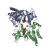
| ||||||||
|---|---|---|---|---|---|---|---|---|---|
| 1 |
| ||||||||
| Unit cell |
|
-Components
| #1: Protein | / FGFR-2 / Keratinocyte growth factor receptor 2 / CD332 antigen Mass: 37038.520 Da / Num. of mol.: 2 / Fragment: Kinase Domain / Mutation: C491A, E565G Source method: isolated from a genetically manipulated source Source: (gene. exp.) Homo sapiens (human) / Gene: FGFR2, BEK, KSAM / Production host:  Escherichia coli (E. coli) / Strain (production host): BL21 (DE3) pLys-S Escherichia coli (E. coli) / Strain (production host): BL21 (DE3) pLys-SReferences: UniProt: P21802, receptor protein-tyrosine kinase#2: Chemical | ChemComp-SO4 / Sulfate  Mass: 96.063 Da / Num. of mol.: 4 / Source method: obtained synthetically / Formula: SO4 Mass: 96.063 Da / Num. of mol.: 4 / Source method: obtained synthetically / Formula: SO4#3: Chemical | ChemComp-MG /   Mass: 24.305 Da / Num. of mol.: 4 / Source method: obtained synthetically / Formula: Mg Mass: 24.305 Da / Num. of mol.: 4 / Source method: obtained synthetically / Formula: Mg#4: Chemical |   Mass: 505.208 Da / Num. of mol.: 2 / Source method: obtained synthetically / Formula: C11H18N5O12P3 / Comment: AMP-PCP, energy-carrying molecule analogue*YM Mass: 505.208 Da / Num. of mol.: 2 / Source method: obtained synthetically / Formula: C11H18N5O12P3 / Comment: AMP-PCP, energy-carrying molecule analogue*YM#5: Water | ChemComp-HOH / | Water Mass: 18.015 Da / Num. of mol.: 172 / Source method: isolated from a natural source / Formula: H2O Mass: 18.015 Da / Num. of mol.: 172 / Source method: isolated from a natural source / Formula: H2O |
|---|
-Experimental details
-Experiment
| Experiment | Method: X-RAY DIFFRACTION / Number of used crystals: 1 |
|---|
- Sample preparation
| Crystal | Density Matthews: 2.62 Å3/Da / Density % sol: 52.99 % |
|---|---|
| Crystal grow | Temperature: 298 K / Method: vapor diffusion, hanging drop / pH: 7.5 Details: 100mM HEPES pH7.5, 25% PEG 4000, 200mM (NH4)2SO4, 2% C3H5(OH)3, VAPOR DIFFUSION, HANGING DROP, temperature 298K |
-Data collection
| Diffraction | Mean temperature: 100 K |
|---|---|
| Diffraction source | Source: SYNCHROTRON / Site: NSLS  / Beamline: X4A / Wavelength: 0.97928 Å / Beamline: X4A / Wavelength: 0.97928 Å |
| Detector | Type: ADSC QUANTUM 4 / Detector: CCD / Date: Sep 17, 2005 |
| Radiation | Monochromator: KOHZU DOUBLE CRYSTAL / Protocol: SINGLE WAVELENGTH / Monochromatic (M) / Laue (L): M / Scattering type: x-ray |
| Radiation wavelength | Wavelength: 0.97928 Å / Relative weight: 1 |
| Reflection | Resolution: 2.3→50 Å / Num. all: 34867 / Num. obs: 34867 / % possible obs: 99.1 % / Observed criterion σ(F): 0 / Observed criterion σ(I): 0 / Redundancy: 5.4 % / Rsym value: 0.064 / Net I/σ(I): 20.3 |
| Reflection shell | Resolution: 2.3→2.38 Å / Redundancy: 3.8 % / Num. unique all: 3243 / Rsym value: 0.226 / % possible all: 93.8 |
- Processing
| Software |
| |||||||||||||||||||||||||
|---|---|---|---|---|---|---|---|---|---|---|---|---|---|---|---|---|---|---|---|---|---|---|---|---|---|---|
| Refinement | Method to determine structure: MOLECULAR REPLACEMENT Starting model: PDB Entry 2PSQ Resolution: 2.3→25 Å / Cross valid method: THROUGHOUT / σ(F): 0 / σ(I): 0 / Stereochemistry target values: Engh & Huber
| |||||||||||||||||||||||||
| Displacement parameters |
| |||||||||||||||||||||||||
| Refinement step | Cycle: LAST / Resolution: 2.3→25 Å
| |||||||||||||||||||||||||
| Refine LS restraints |
|
