 Movie
Movie Controller

[English] 日本語
 Yorodumi
- PDB-2px7: Crystal structure of 2-C-methyl-D-erythritol 4-phosphate cytidyly... -
+ Open data

- Basic information
| Entry | Database: PDB / ID: 2px7 | ||||||
|---|---|---|---|---|---|---|---|
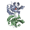
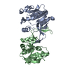
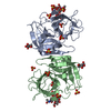
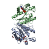
| Title | Crystal structure of 2-C-methyl-D-erythritol 4-phosphate cytidylyltransferase from Thermus thermophilus HB8 | ||||||
 Components Components | 2-C-methyl-D-erythritol 4-phosphate cytidylyltransferase | ||||||
 Keywords Keywords | TRANSFERASE / ttha0171 / 2-C-methyl-D-erythritol 4-phosphate cytidylyltransferase / ISPD_THET8 / ispD / Structural genomics / PSI / Protein Structure Initiative / Southeast Collaboratory for Structural Genomics / SECSG / NPPSFA / National Project on Protein Structural and Functional Analyses / RIKEN STRUCTURAL GENOMICS/PROTEOMICS INITIATIVE / RSGI | ||||||
| Function / homology |  Function and homology information2-C-methyl-D-erythritol 4-phosphate cytidylyltransferase / 2-C-methyl-D-erythritol 4-phosphate cytidylyltransferase activity / isopentenyl diphosphate biosynthetic process, methylerythritol 4-phosphate pathway / terpenoid biosynthetic process Function and homology information2-C-methyl-D-erythritol 4-phosphate cytidylyltransferase / 2-C-methyl-D-erythritol 4-phosphate cytidylyltransferase activity / isopentenyl diphosphate biosynthetic process, methylerythritol 4-phosphate pathway / terpenoid biosynthetic processSimilarity search - Function | ||||||
| Biological species |   Thermus thermophilus HB8 (bacteria) Thermus thermophilus HB8 (bacteria) | ||||||
| Method | X-RAY DIFFRACTION / SYNCHROTRON / MOLECULAR REPLACEMENT / Resolution: 2.2 Å | ||||||
 Authors Authors | Chen, L. / Tsukuda, M. / Ebihara, A. / Shinkai, A. / Kuramitsu, S. / Yokoyama, S. / Chen, L.-Q. / Liu, Z.-J. / Lee, D. / Chang, S.-H. ...Chen, L. / Tsukuda, M. / Ebihara, A. / Shinkai, A. / Kuramitsu, S. / Yokoyama, S. / Chen, L.-Q. / Liu, Z.-J. / Lee, D. / Chang, S.-H. / Nguyen, D. / Rose, J.P. / Wang, B.-C. / Southeast Collaboratory for Structural Genomics (SECSG) / RIKEN Structural Genomics/Proteomics Initiative (RSGI) | ||||||
 Citation Citation | Journal: To be Published Title: Crystal structure of 2-C-methyl-D-erythritol 4-phosphate cytidylyltransferase from Thermus thermophilus HB8. Authors: Chen, L. / Tsukuda, M. / Ebihara, A. / Shinkai, A. / Kuramitsu, S. / Yokoyama, S. / Chen, L.-Q. / Liu, Z.-J. / Lee, D. / Chang, S.-H. / Nguyen, D. / Rose, J.P. / Wang, B.-C. | ||||||
| History |
|
- Structure visualization
| Structure viewer | Molecule: MolmilJmol/JSmol |
|---|
- Downloads & links
-Download
| PDBx/mmCIF format | 2px7.cif.gz | 92 KB | Display | PDBx/mmCIF format |
|---|---|---|---|---|
| PDB format | pdb2px7.ent.gz | 69.3 KB | Display | PDB format |
| PDBx/mmJSON format | 2px7.json.gz | Tree view | PDBx/mmJSON format | |
| Others |  Other downloads Other downloads |
-Validation report
| Arichive directory | https://data.pdbj.org/pub/pdb/validation_reports/px/2px7ftp://data.pdbj.org/pub/pdb/validation_reports/px/2px7 | HTTPS FTP |
|---|
-Related structure data
| Related structure data |  1ye7 S: Starting model for refinement |
|---|---|
| Similar structure data | |
| Other databases |
|


-Links
 PDBj
PDBj

- Assembly
| Deposited unit | 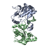
| ||||||||
|---|---|---|---|---|---|---|---|---|---|
| 1 |
| ||||||||
| Unit cell |
|
-Components
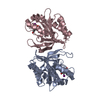
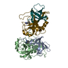
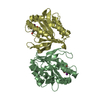
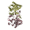
| #1: Protein | / 4-diphosphocytidyl-2C-methyl-D-erythritol synthase / MEP cytidylyltransferase / MCT Mass: 25226.848 Da / Num. of mol.: 2 Source method: isolated from a genetically manipulated source Source: (gene. exp.) Thermus thermophilus HB8 (bacteria) / Species: Thermus thermophilus / Strain: HB8, DSM 579 / Gene: ispD, TTHA0171 / Plasmid: pET-19b / Production host: Escherichia coli (E. coli) / Strain (production host): Rosetta(DE3)References: UniProt: Q5SLX2, 2-C-methyl-D-erythritol 4-phosphate cytidylyltransferase#2: Water | ChemComp-HOH / | Water Mass: 18.015 Da / Num. of mol.: 160 / Source method: isolated from a natural source / Formula: H2O Mass: 18.015 Da / Num. of mol.: 160 / Source method: isolated from a natural source / Formula: H2O |
|---|
-Experimental details
-Experiment
| Experiment | Method: X-RAY DIFFRACTION / Number of used crystals: 1 |
|---|
- Sample preparation
| Crystal | Density Matthews: 3.34 Å3/Da / Density % sol: 63.15 % |
|---|---|
| Crystal grow | Temperature: 293 K / Method: vapor diffusion, sitting drop / pH: 8 Details: USING 2 MICROLITER DROPS CONTAINING EQUAL VOLUMES OF PROTEIN CONCENTRATE (6.36 mg/ml) AND RESERVOIR SOLUTION CONTAINING 0.9M Li Sulfate, 0.09M HEPES pH 8.0, 0.01M Betaine, VAPOR DIFFUSION, ...Details: USING 2 MICROLITER DROPS CONTAINING EQUAL VOLUMES OF PROTEIN CONCENTRATE (6.36 mg/ml) AND RESERVOIR SOLUTION CONTAINING 0.9M Li Sulfate, 0.09M HEPES pH 8.0, 0.01M Betaine, VAPOR DIFFUSION, SITTING DROP, temperature 293K |
-Data collection
| Diffraction | Mean temperature: 100 K |
|---|---|
| Diffraction source | Source: SYNCHROTRON / Site: SPring-8  / Beamline: BL26B2 / Wavelength: 1 Å / Beamline: BL26B2 / Wavelength: 1 Å |
| Detector | Type: RIGAKU JUPITER 210 / Detector: CCD / Date: Mar 28, 2007 |
| Radiation | Protocol: SINGLE WAVELENGTH / Monochromatic (M) / Laue (L): M / Scattering type: x-ray |
| Radiation wavelength | Wavelength: 1 Å / Relative weight: 1 |
| Reflection | Resolution: 2.2→50 Å / Num. obs: 33616 / % possible obs: 100 % / Redundancy: 11 % / Rsym value: 0.043 / Χ2: 0.942 / Net I/σ(I): 25.6 |
| Reflection shell | Resolution: 2.2→2.28 Å / Redundancy: 11 % / Num. unique all: 3339 / Rsym value: 0.286 / Χ2: 1.071 / % possible all: 100 |
- Processing
| Software |
| ||||||||||||||||||||||||||||||||||||||||||||||||||||||||||||||||||||||||||||||||||||||||||
|---|---|---|---|---|---|---|---|---|---|---|---|---|---|---|---|---|---|---|---|---|---|---|---|---|---|---|---|---|---|---|---|---|---|---|---|---|---|---|---|---|---|---|---|---|---|---|---|---|---|---|---|---|---|---|---|---|---|---|---|---|---|---|---|---|---|---|---|---|---|---|---|---|---|---|---|---|---|---|---|---|---|---|---|---|---|---|---|---|---|---|---|
| Refinement | Method to determine structure: MOLECULAR REPLACEMENT Starting model: PDB entry 1YE7 1ye7 Resolution: 2.2→50 Å / Cor.coef. Fo:Fc: 0.935 / Cor.coef. Fo:Fc free: 0.921 / Cross valid method: THROUGHOUT / σ(F): 0 / ESU R: 0.206 / ESU R Free: 0.175 / Stereochemistry target values: MAXIMUM LIKELIHOOD / Details: HYDROGENS HAVE BEEN ADDED IN THE RIDING POSITIONS
| ||||||||||||||||||||||||||||||||||||||||||||||||||||||||||||||||||||||||||||||||||||||||||
| Solvent computation | Ion probe radii: 0.8 Å / Shrinkage radii: 0.8 Å / VDW probe radii: 1.4 Å / Solvent model: MASK | ||||||||||||||||||||||||||||||||||||||||||||||||||||||||||||||||||||||||||||||||||||||||||
| Displacement parameters | Biso mean: 37.878 Å2
| ||||||||||||||||||||||||||||||||||||||||||||||||||||||||||||||||||||||||||||||||||||||||||
| Refinement step | Cycle: LAST / Resolution: 2.2→50 Å
| ||||||||||||||||||||||||||||||||||||||||||||||||||||||||||||||||||||||||||||||||||||||||||
| Refine LS restraints |
| ||||||||||||||||||||||||||||||||||||||||||||||||||||||||||||||||||||||||||||||||||||||||||
| LS refinement shell | Resolution: 2.2→2.258 Å / Total num. of bins used: 20
|
